 Movie
Movie Controller

[English] 日本語
 Yorodumi
- PDB-8xll: Structure of the native 2-oxoglutarate dehydrogenase complex (OGD... -
+ Open data

- Basic information
| Entry | Database: PDB / ID: 8xll | |||||||||||||||||||||||||||||||||||||||||||||
|---|---|---|---|---|---|---|---|---|---|---|---|---|---|---|---|---|---|---|---|---|---|---|---|---|---|---|---|---|---|---|---|---|---|---|---|---|---|---|---|---|---|---|---|---|---|---|
| Title | Structure of the native 2-oxoglutarate dehydrogenase complex (OGDHC) in the adult cortex and hippocampus | |||||||||||||||||||||||||||||||||||||||||||||
 Components Components | Dihydrolipoyllysine-residue succinyltransferase component of 2-oxoglutarate dehydrogenase complex, mitochondrial | |||||||||||||||||||||||||||||||||||||||||||||
 Keywords Keywords | TRANSFERASE / 2-oxoglutarate dehydrogenase complex (OGDHC) | |||||||||||||||||||||||||||||||||||||||||||||
| Function / homology |  Function and homology information Function and homology informationGlycine degradation / OGDH complex synthesizes succinyl-CoA from 2-OG / Protein lipoylation / OADH complex synthesizes glutaryl-CoA from 2-OA / 2-oxoglutarate decarboxylation to succinyl-CoA / oxoadipate dehydrogenase complex / : / succinyl-CoA metabolic process / dihydrolipoyllysine-residue succinyltransferase / dihydrolipoyllysine-residue succinyltransferase activity ...Glycine degradation / OGDH complex synthesizes succinyl-CoA from 2-OG / Protein lipoylation / OADH complex synthesizes glutaryl-CoA from 2-OA / 2-oxoglutarate decarboxylation to succinyl-CoA / oxoadipate dehydrogenase complex / : / succinyl-CoA metabolic process / dihydrolipoyllysine-residue succinyltransferase / dihydrolipoyllysine-residue succinyltransferase activity / oxoglutarate dehydrogenase complex / 2-oxoglutarate metabolic process / acyltransferase activity / tricarboxylic acid cycle / heat shock protein binding / sarcolemma / protein-folding chaperone binding / mitochondrial matrix / mitochondrion / nucleoplasm / nucleus Similarity search - Function | |||||||||||||||||||||||||||||||||||||||||||||
| Biological species |  | |||||||||||||||||||||||||||||||||||||||||||||
| Method | ELECTRON MICROSCOPY / single particle reconstruction / cryo EM / Resolution: 3.1 Å | |||||||||||||||||||||||||||||||||||||||||||||
 Authors Authors | Zhang, M. / Feng, J. / Li, Y. / Zhu, S. | |||||||||||||||||||||||||||||||||||||||||||||
| Funding support |  China, 1items China, 1items
| |||||||||||||||||||||||||||||||||||||||||||||
 Citation Citation | Journal: Cell / Year: 2025 Title: Assembly and architecture of endogenous NMDA receptors in adult cerebral cortex and hippocampus. Authors: Ming Zhang / Juan Feng / Chun Xie / Nan Song / Chaozhi Jin / Jian Wang / Qun Zhao / Lihua Zhang / Boshuang Wang / Yidi Sun / Fei Guo / Yang Li / Shujia Zhu / Abstract: The cerebral cortex and hippocampus are crucial brain regions for learning and memory, which depend on activity-induced synaptic plasticity involving N-methyl-ᴅ-aspartate receptors (NMDARs). ...The cerebral cortex and hippocampus are crucial brain regions for learning and memory, which depend on activity-induced synaptic plasticity involving N-methyl-ᴅ-aspartate receptors (NMDARs). However, subunit assembly and molecular architecture of endogenous NMDARs (eNMDARs) in the brain remain elusive. Using conformation- and subunit-dependent antibodies, we purified eNMDARs from adult rat cerebral cortex and hippocampus. Three major subtypes of GluN1-N2A-N2B, GluN1-N2B, and GluN1-N2A eNMDARs were resolved by cryoelectron microscopy (cryo-EM) at the resolution up to 4.2 Å. The particle ratio of these three subtypes was 9:7:4, indicating that about half of GluN2A and GluN2B subunits are incorporated into the tri-heterotetramers. Structural analysis revealed the asymmetric architecture of the GluN1-N2A-N2B receptor throughout the extracellular to the transmembrane layers. Moreover, the conformational variations between GluN1-N2B and GluN1-N2A-N2B receptors revealed the distinct biophysical properties across different eNMDAR subtypes. Our findings imply the structural and functional complexity of eNMDARs and shed light on structure-based therapeutic design targeting these eNMDARs in vivo. | |||||||||||||||||||||||||||||||||||||||||||||
| History |
|
- Structure visualization
| Structure viewer | Molecule: MolmilJmol/JSmol |
|---|
- Downloads & links
-Download
| PDBx/mmCIF format | 8xll.cif.gz | 927.3 KB | Display | PDBx/mmCIF format |
|---|---|---|---|---|
| PDB format | pdb8xll.ent.gz | 785.3 KB | Display | PDB format |
| PDBx/mmJSON format | 8xll.json.gz | Tree view | PDBx/mmJSON format | |
| Others |  Other downloads Other downloads |
-Validation report
| Arichive directory | https://data.pdbj.org/pub/pdb/validation_reports/xl/8xllftp://data.pdbj.org/pub/pdb/validation_reports/xl/8xll | HTTPS FTP |
|---|
-Related structure data
| Related structure data |  38452MC  8xljC 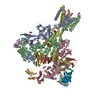 8xlkC  9jnnC M: map data used to model this data C: citing same article ( |
|---|---|
| Similar structure data |


-Links
 PDBj
PDBj

- Assembly
| Deposited unit | 
|
|---|---|
| 1 |
|
-Components
| #1: Protein | Mass: 23953.686 Da / Num. of mol.: 24 / Source method: isolated from a natural source / Source: (natural) References: UniProt: Q01205, dihydrolipoyllysine-residue succinyltransferase Has protein modification | N | |
|---|
-Experimental details
-Experiment
| Experiment | Method: ELECTRON MICROSCOPY |
|---|---|
| EM experiment | Aggregation state: TISSUE / 3D reconstruction method: single particle reconstruction |
- Sample preparation
| Component | Name: Mammalian OGDHC from rat brain / Type: COMPLEX / Entity ID: all / Source: NATURAL |
|---|---|
| Molecular weight | Units: MEGADALTONS / Experimental value: NO |
| Source (natural) | Organism: |
| Buffer solution | pH: 8 |
| Specimen | Embedding applied: NO / Shadowing applied: NO / Staining applied: NO / Vitrification applied: YES |
| Vitrification | Cryogen name: ETHANE |
- Electron microscopy imaging
| Experimental equipment |  Model: Titan Krios / Image courtesy: FEI Company |
|---|---|
| Microscopy | Model: FEI TITAN KRIOS |
| Electron gun | Electron source:  FIELD EMISSION GUN / Accelerating voltage: 300 kV / Illumination mode: FLOOD BEAM FIELD EMISSION GUN / Accelerating voltage: 300 kV / Illumination mode: FLOOD BEAM |
| Electron lens | Mode: BRIGHT FIELD / Nominal defocus max: 2500 nm / Nominal defocus min: 1000 nm |
| Image recording | Electron dose: 60 e/Å2 / Film or detector model: DIRECT ELECTRON DE-10 (5k x 4k) |
- Processing
| EM software | Name: PHENIX / Category: model refinement | ||||||||||||||||||||||||
|---|---|---|---|---|---|---|---|---|---|---|---|---|---|---|---|---|---|---|---|---|---|---|---|---|---|
| CTF correction | Type: NONE | ||||||||||||||||||||||||
| 3D reconstruction | Resolution: 3.1 Å / Resolution method: FSC 0.143 CUT-OFF / Num. of particles: 268960 / Symmetry type: POINT | ||||||||||||||||||||||||
| Refine LS restraints |
|
