[English] 日本語
Yorodumi
- EMDB-38452: Structure of the native 2-oxoglutarate dehydrogenase complex (OGD... -

+
Open data


ID or keywords:

Loading...

-
Basic information

Entry
Database: EMDB / ID: EMD-38452
TitleStructure of the native 2-oxoglutarate dehydrogenase complex (OGDHC) in the adult cortex and hippocampus
Map data
Sample
  • Complex: Mammalian OGDHC from rat brain
    • Protein or peptide: Dihydrolipoyllysine-residue succinyltransferase component of 2-oxoglutarate dehydrogenase complex, mitochondrial
Keywords2-oxoglutarate dehydrogenase complex (OGDHC) / TRANSFERASE
Function / homology
Function and homology information


Glycine degradation / OGDH complex synthesizes succinyl-CoA from 2-OG / Protein lipoylation / OADH complex synthesizes glutaryl-CoA from 2-OA / oxoadipate dehydrogenase complex / L-lysine catabolic process to acetyl-CoA via saccharopine / succinyl-CoA metabolic process / dihydrolipoyllysine-residue succinyltransferase / dihydrolipoyllysine-residue succinyltransferase activity / oxoglutarate dehydrogenase complex ...Glycine degradation / OGDH complex synthesizes succinyl-CoA from 2-OG / Protein lipoylation / OADH complex synthesizes glutaryl-CoA from 2-OA / oxoadipate dehydrogenase complex / L-lysine catabolic process to acetyl-CoA via saccharopine / succinyl-CoA metabolic process / dihydrolipoyllysine-residue succinyltransferase / dihydrolipoyllysine-residue succinyltransferase activity / oxoglutarate dehydrogenase complex / 2-oxoglutarate metabolic process / : / acyltransferase activity / tricarboxylic acid cycle / heat shock protein binding / protein-folding chaperone binding / mitochondrial matrix / mitochondrion / nucleus
Similarity search - Function
: / Dihydrolipoamide succinyltransferase / 2-oxo acid dehydrogenase, lipoyl-binding site / 2-oxo acid dehydrogenases acyltransferase component lipoyl binding site. / 2-oxoacid dehydrogenase acyltransferase, catalytic domain / 2-oxoacid dehydrogenases acyltransferase (catalytic domain) / Biotin-requiring enzyme / Biotinyl/lipoyl domain profile. / Biotin/lipoyl attachment / Single hybrid motif / Chloramphenicol acetyltransferase-like domain superfamily
Similarity search - Domain/homology
Dihydrolipoyllysine-residue succinyltransferase component of 2-oxoglutarate dehydrogenase complex, mitochondrial
Similarity search - Component
Biological speciesRattus norvegicus (Norway rat)
Methodsingle particle reconstruction / cryo EM / Resolution: 3.1 Å
AuthorsZhang M / Feng J / Li Y / Zhu S
Funding support China, 1 items
OrganizationGrant numberCountry
Chinese Academy of Sciences China
Citation
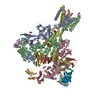
Journal: Cell / Year: 2025
Title: Assembly and architecture of endogenous NMDA receptors in adult cerebral cortex and hippocampus.
Authors: Ming Zhang / Juan Feng / Chun Xie / Nan Song / Chaozhi Jin / Jian Wang / Qun Zhao / Lihua Zhang / Boshuang Wang / Yidi Sun / Fei Guo / Yang Li / Shujia Zhu /
Abstract: The cerebral cortex and hippocampus are crucial brain regions for learning and memory, which depend on activity-induced synaptic plasticity involving N-methyl-ᴅ-aspartate receptors (NMDARs). ...The cerebral cortex and hippocampus are crucial brain regions for learning and memory, which depend on activity-induced synaptic plasticity involving N-methyl-ᴅ-aspartate receptors (NMDARs). However, subunit assembly and molecular architecture of endogenous NMDARs (eNMDARs) in the brain remain elusive. Using conformation- and subunit-dependent antibodies, we purified eNMDARs from adult rat cerebral cortex and hippocampus. Three major subtypes of GluN1-N2A-N2B, GluN1-N2B, and GluN1-N2A eNMDARs were resolved by cryoelectron microscopy (cryo-EM) at the resolution up to 4.2 Å. The particle ratio of these three subtypes was 9:7:4, indicating that about half of GluN2A and GluN2B subunits are incorporated into the tri-heterotetramers. Structural analysis revealed the asymmetric architecture of the GluN1-N2A-N2B receptor throughout the extracellular to the transmembrane layers. Moreover, the conformational variations between GluN1-N2B and GluN1-N2A-N2B receptors revealed the distinct biophysical properties across different eNMDAR subtypes. Our findings imply the structural and functional complexity of eNMDARs and shed light on structure-based therapeutic design targeting these eNMDARs in vivo.
History
DepositionDec 26, 2023-
Header (metadata) releaseFeb 5, 2025-
Map releaseFeb 5, 2025-
UpdateMar 19, 2025-
Current statusMar 19, 2025Processing site: PDBj / Status: Released

-
Structure visualization

Supplemental images

Downloads & links

-
Map

FileDownload / File: emd_38452.map.gz / Format: CCP4 / Size: 303.3 MB / Type: IMAGE STORED AS FLOATING POINT NUMBER (4 BYTES)
Projections & slices

Image control

Size
Brightness
Contrast
Others
AxesZ (Sec.)Y (Row.)X (Col.)
1.07 Å/pix.
x 430 pix.
= 460.53 Å
1.07 Å/pix.
x 430 pix.
= 460.53 Å
1.07 Å/pix.
x 430 pix.
= 460.53 Å

Surface

Projections

Slices (1/3)

Slices (1/2)

Slices (2/3)

Images are generated by Spider.

Voxel sizeX=Y=Z: 1.071 Å
Density
Contour LevelBy AUTHOR: 0.288
Minimum - Maximum-0.9332667 - 1.418375
Average (Standard dev.)0.0005567981 (±0.033522394)
SymmetrySpace group: 1
Details

EMDB XML:

Map geometry
Axis orderXYZ
Origin000
Dimensions430430430
Spacing430430430
CellA=B=C: 460.53 Å
α=β=γ: 90.0 °

-
Supplemental data

-
Mask #1

Fileemd_38452_msk_1.map
Projections & Slices
AxesZYX

Projections

Slices (1/2)
Density Histograms

-
Half map: #2

Fileemd_38452_half_map_1.map
Projections & Slices
AxesZYX

Projections

Slices (1/2)
Density Histograms

-
Half map: #1

Fileemd_38452_half_map_2.map
Projections & Slices
AxesZYX

Projections

Slices (1/2)
Density Histograms

-
Sample components

-
Entire : Mammalian OGDHC from rat brain

EntireName: Mammalian OGDHC from rat brain
Components
  • Complex: Mammalian OGDHC from rat brain
    • Protein or peptide: Dihydrolipoyllysine-residue succinyltransferase component of 2-oxoglutarate dehydrogenase complex, mitochondrial

-
Supramolecule #1: Mammalian OGDHC from rat brain

SupramoleculeName: Mammalian OGDHC from rat brain / type: complex / ID: 1 / Parent: 0 / Macromolecule list: all
Source (natural)Organism: Rattus norvegicus (Norway rat)

-
Macromolecule #1: Dihydrolipoyllysine-residue succinyltransferase component of 2-ox...

MacromoleculeName: Dihydrolipoyllysine-residue succinyltransferase component of 2-oxoglutarate dehydrogenase complex, mitochondrial
type: protein_or_peptide / ID: 1 / Number of copies: 24 / Enantiomer: LEVO / EC number: dihydrolipoyllysine-residue succinyltransferase
Source (natural)Organism: Rattus norvegicus (Norway rat)
Molecular weightTheoretical: 23.953686 KDa
SequenceString: LKEAQNTCAM LTTFNEVDMS NIQEMRARHK DAFLKKHNLK LGFMSAFVKA SAFALQEQPV VNAVIDDATK EVVYRDYIDI SVAVATPRG LVVPVIRNVE TMNYADIERT INELGEKARK NELAIEDMDG GTFTISNGGV FGSLFGTPII NPPQSAILGM H GIFDRPVA ...String:
LKEAQNTCAM LTTFNEVDMS NIQEMRARHK DAFLKKHNLK LGFMSAFVKA SAFALQEQPV VNAVIDDATK EVVYRDYIDI SVAVATPRG LVVPVIRNVE TMNYADIERT INELGEKARK NELAIEDMDG GTFTISNGGV FGSLFGTPII NPPQSAILGM H GIFDRPVA VGGKVEVRPM MYVALTYDHR LIDGREAVTF LRKIKAAVED PRVLLLDL

UniProtKB: Dihydrolipoyllysine-residue succinyltransferase component of 2-oxoglutarate dehydrogenase complex, mitochondrial

-
Experimental details

-
Structure determination

Methodcryo EM
Processingsingle particle reconstruction
Aggregation statetissue

-
Sample preparation

BufferpH: 8
VitrificationCryogen name: ETHANE

-
Electron microscopy

MicroscopeFEI TITAN KRIOS
Image recordingFilm or detector model: DIRECT ELECTRON DE-10 (5k x 4k) / Average electron dose: 60.0 e/Å2
Electron beamAcceleration voltage: 300 kV / Electron source: FIELD EMISSION GUN
Electron opticsIllumination mode: FLOOD BEAM / Imaging mode: BRIGHT FIELD / Nominal defocus max: 2.5 µm / Nominal defocus min: 1.0 µm
Experimental equipment
Model: Titan Krios / Image courtesy: FEI Company

+
Image processing

Startup modelType of model: OTHER
Final reconstructionResolution.type: BY AUTHOR / Resolution: 3.1 Å / Resolution method: FSC 0.143 CUT-OFF / Number images used: 268960
Initial angle assignmentType: OTHER
Final angle assignmentType: OTHER
FSC plot (resolution estimation)

+
About Yorodumi

-
News

-
Feb 9, 2022. New format data for meta-information of EMDB entries

New format data for meta-information of EMDB entries

  • Version 3 of the EMDB header file is now the official format.
  • The previous official version 1.9 will be removed from the archive.

Related info.:EMDB header

External links:wwPDB to switch to version 3 of the EMDB data model

-
Aug 12, 2020. Covid-19 info

Covid-19 info

URL: https://pdbj.org/emnavi/covid19.php

New page: Covid-19 featured information page in EM Navigator.

Related info.:Covid-19 info / Mar 5, 2020. Novel coronavirus structure data

+
Mar 5, 2020. Novel coronavirus structure data

Novel coronavirus structure data

Related info.:Yorodumi Speices / Aug 12, 2020. Covid-19 info

External links:COVID-19 featured content - PDBj / Molecule of the Month (242):Coronavirus Proteases

+
Jan 31, 2019. EMDB accession codes are about to change! (news from PDBe EMDB page)

EMDB accession codes are about to change! (news from PDBe EMDB page)

  • The allocation of 4 digits for EMDB accession codes will soon come to an end. Whilst these codes will remain in use, new EMDB accession codes will include an additional digit and will expand incrementally as the available range of codes is exhausted. The current 4-digit format prefixed with “EMD-” (i.e. EMD-XXXX) will advance to a 5-digit format (i.e. EMD-XXXXX), and so on. It is currently estimated that the 4-digit codes will be depleted around Spring 2019, at which point the 5-digit format will come into force.
  • The EM Navigator/Yorodumi systems omit the EMD- prefix.

Related info.:Q: What is EMD? / ID/Accession-code notation in Yorodumi/EM Navigator

External links:EMDB Accession Codes are Changing Soon! / Contact to PDBj

+
Jul 12, 2017. Major update of PDB

Major update of PDB

  • wwPDB released updated PDB data conforming to the new PDBx/mmCIF dictionary.
  • This is a major update changing the version number from 4 to 5, and with Remediation, in which all the entries are updated.
  • In this update, many items about electron microscopy experimental information are reorganized (e.g. em_software).
  • Now, EM Navigator and Yorodumi are based on the updated data.

External links:wwPDB Remediation / Enriched Model Files Conforming to OneDep Data Standards Now Available in the PDB FTP Archive

-
Yorodumi

Thousand views of thousand structures

  • Yorodumi is a browser for structure data from EMDB, PDB, SASBDB, etc.
  • This page is also the successor to EM Navigator detail page, and also detail information page/front-end page for Omokage search.
  • The word "yorodu" (or yorozu) is an old Japanese word meaning "ten thousand". "mi" (miru) is to see.

Related info.:EMDB / PDB / SASBDB / Comparison of 3 databanks / Yorodumi Search / Aug 31, 2016. New EM Navigator & Yorodumi / Yorodumi Papers / Jmol/JSmol / Function and homology information / Changes in new EM Navigator and Yorodumi

Read more