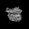
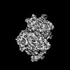
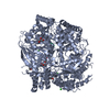
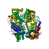
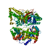
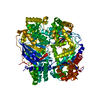
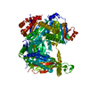
[English] 日本語
Yorodumi
- PDB-8wxz: Falcilysin in complex with hemoglobin beta chain peptide -

+
Open data


ID or keywords:

Loading...

-
Basic information

Entry
Database: PDB / ID: 8wxz
TitleFalcilysin in complex with hemoglobin beta chain peptide
Components
  • Falcilysin
  • Hemoglobin subunit beta fragment
KeywordsHYDROLASE / falcilysin-substrate complex / inactive mutant
Function / homology
Function and homology information


hemoglobin catabolic process / apicoplast / food vacuole / Hydrolases; Acting on peptide bonds (peptidases); Metalloendopeptidases / vacuolar membrane / nitric oxide transport / cellular oxidant detoxification / hemoglobin binding / hemoglobin alpha binding / haptoglobin-hemoglobin complex ...hemoglobin catabolic process / apicoplast / food vacuole / Hydrolases; Acting on peptide bonds (peptidases); Metalloendopeptidases / vacuolar membrane / nitric oxide transport / cellular oxidant detoxification / hemoglobin binding / hemoglobin alpha binding / haptoglobin-hemoglobin complex / organic acid binding / renal absorption / hemoglobin complex / oxygen transport / Scavenging of heme from plasma / endocytic vesicle lumen / blood vessel diameter maintenance / hydrogen peroxide catabolic process / oxygen carrier activity / Late endosomal microautophagy / Heme signaling / carbon dioxide transport / Erythrocytes take up oxygen and release carbon dioxide / response to hydrogen peroxide / Erythrocytes take up carbon dioxide and release oxygen / Cytoprotection by HMOX1 / protein processing / metalloendopeptidase activity / oxygen binding / platelet aggregation / regulation of blood pressure / Chaperone Mediated Autophagy / positive regulation of nitric oxide biosynthetic process / tertiary granule lumen / Factors involved in megakaryocyte development and platelet production / ficolin-1-rich granule lumen / blood microparticle / heme binding / Neutrophil degranulation / extracellular space / extracellular exosome / extracellular region / metal ion binding / cytosol
Similarity search - Function
Peptidase M16C associated / Peptidase M16C associated / Peptidase M16C associated / Peptidase M16, C-terminal / Peptidase M16 inactive domain / Peptidase M16, N-terminal / Insulinase (Peptidase family M16) / : / Metalloenzyme, LuxS/M16 peptidase-like / Hemoglobin, beta-type ...Peptidase M16C associated / Peptidase M16C associated / Peptidase M16C associated / Peptidase M16, C-terminal / Peptidase M16 inactive domain / Peptidase M16, N-terminal / Insulinase (Peptidase family M16) / : / Metalloenzyme, LuxS/M16 peptidase-like / Hemoglobin, beta-type / Globin/Protoglobin / Globin domain profile. / Globin / Globin / Globin-like superfamily
Similarity search - Domain/homology
ACETIC ACID / DI(HYDROXYETHYL)ETHER / Hemoglobin subunit beta / Falcilysin
Similarity search - Component
Biological speciesPlasmodium falciparum 3D7 (eukaryote)
Homo sapiens (human)
MethodX-RAY DIFFRACTION / SYNCHROTRON / MOLECULAR REPLACEMENT / Resolution: 2.3 Å
AuthorsLin, J.Q. / Lescar, J.
Funding support1items
OrganizationGrant numberCountry
Not funded
CitationJournal: To Be Published
Title: Falcilysin in complex with hemoglobin alpha chain peptide
Authors: Lin, J.Q. / Lescar, J.
History
DepositionOct 30, 2023Deposition site: PDBJ / Processing site: PDBJ
Revision 1.0Aug 7, 2024Provider: repository / Type: Initial release

-
Structure visualization

Structure viewerMolecule:
MolmilJmol/JSmol

Downloads & links

-
Assembly

Deposited unit
A: Falcilysin
P: Hemoglobin subunit beta fragment
hetero molecules


Theoretical massNumber of molelcules
Total (without water)137,1495
Polymers136,9172
Non-polymers2323
Water5,423301
1


  • Idetical with deposited unit
  • defined by author&software
  • Evidence: electron microscopy
TypeNameSymmetry operationNumber
identity operation1_555x,y,z1
Buried area1760 Å2
ΔGint-43 kcal/mol
Surface area42150 Å2
MethodPISA
Unit cell
Length a, b, c (Å)94.090, 105.870, 114.910
Angle α, β, γ (deg.)90.000, 90.000, 90.000
Int Tables number19
Space group name H-MP212121
Space group name HallP2ac2ab
Symmetry operation#1: x,y,z
#2: x+1/2,-y+1/2,-z
#3: -x,y+1/2,-z+1/2
#4: -x+1/2,-y,z+1/2

-
Components

-
Protein / Protein/peptide , 2 types, 2 molecules AP

#1: Protein Falcilysin


Mass: 135038.375 Da / Num. of mol.: 1 / Mutation: E132Q
Source method: isolated from a genetically manipulated source
Source: (gene. exp.) Plasmodium falciparum 3D7 (eukaryote) / Gene: FLN, PF3D7_1360800 / Production host: Escherichia coli (E. coli)
References: UniProt: Q76NL8, Hydrolases; Acting on peptide bonds (peptidases); Metalloendopeptidases
#2: Protein/peptide Hemoglobin subunit beta fragment


Mass: 1879.054 Da / Num. of mol.: 1 / Source method: obtained synthetically / Source: (synth.) Homo sapiens (human) / References: UniProt: P68871

-
Non-polymers , 4 types, 304 molecules

#3: Chemical ChemComp-PEG / DI(HYDROXYETHYL)ETHER


Mass: 106.120 Da / Num. of mol.: 1 / Source method: obtained synthetically / Formula: C4H10O3
#4: Chemical ChemComp-ACY / ACETIC ACID


Mass: 60.052 Da / Num. of mol.: 1 / Source method: obtained synthetically / Formula: C2H4O2
#5: Chemical ChemComp-ZN / ZINC ION


Mass: 65.409 Da / Num. of mol.: 1 / Source method: obtained synthetically / Formula: Zn / Feature type: SUBJECT OF INVESTIGATION
#6: Water ChemComp-HOH / water


Mass: 18.015 Da / Num. of mol.: 301 / Source method: isolated from a natural source / Formula: H2O

-
Details

Has ligand of interestY

-
Experimental details

-
Experiment

ExperimentMethod: X-RAY DIFFRACTION / Number of used crystals: 1

-
Sample preparation

CrystalDensity Matthews: 2.09 Å3/Da / Density % sol: 41.15 %
Crystal growTemperature: 293.15 K / Method: vapor diffusion, sitting drop
Details: 0.12M monosaccharides, 0.1M imidazole/MES monohydrate pH 6.5, 20% v/v PEG 500, 10% w/v PEG 20000

-
Data collection

DiffractionMean temperature: 100 K / Serial crystal experiment: N
Diffraction sourceSource: SYNCHROTRON / Site: Australian Synchrotron / Beamline: MX2 / Wavelength: 0.9537 Å
DetectorType: ADSC QUANTUM 315r / Detector: CCD / Date: Mar 2, 2022
RadiationProtocol: SINGLE WAVELENGTH / Monochromatic (M) / Laue (L): M / Scattering type: x-ray
Radiation wavelengthWavelength: 0.9537 Å / Relative weight: 1
ReflectionResolution: 2.3→48.08 Å / Num. obs: 51607 / % possible obs: 99.91 % / Redundancy: 7.4 % / Biso Wilson estimate: 36.89 Å2 / CC1/2: 0.996 / CC star: 0.999 / Rmerge(I) obs: 0.1428 / Net I/σ(I): 10.3
Reflection shellResolution: 2.3→2.382 Å / Redundancy: 7.1 % / Num. unique obs: 5041 / CC1/2: 0.748 / CC star: 0.925 / % possible all: 99.9

-
Processing

Software
NameVersionClassification
PHENIX1.21.1-5286-0000refinement
XDSdata reduction
XDSdata scaling
Cootmodel building
PHASERphasing
RefinementMethod to determine structure: MOLECULAR REPLACEMENT / Resolution: 2.3→48.08 Å / SU ML: 0.2779 / Cross valid method: FREE R-VALUE / σ(F): 0.98 / Phase error: 25.6835
Stereochemistry target values: GeoStd + Monomer Library + CDL v1.2
RfactorNum. reflection% reflection
Rfree0.2505 4905 4.99 %
Rwork0.2048 93426 -
obs0.2071 51597 99.86 %
Solvent computationShrinkage radii: 0.9 Å / VDW probe radii: 1.11 Å / Solvent model: FLAT BULK SOLVENT MODEL
Displacement parametersBiso mean: 41.73 Å2
Refinement stepCycle: LAST / Resolution: 2.3→48.08 Å
ProteinNucleic acidLigandSolventTotal
Num. atoms8944 0 12 301 9257
Refine LS restraints
Refine-IDTypeDev idealNumber
X-RAY DIFFRACTIONf_bond_d0.00569133
X-RAY DIFFRACTIONf_angle_d0.811412292
X-RAY DIFFRACTIONf_chiral_restr0.04851340
X-RAY DIFFRACTIONf_plane_restr0.00421569
X-RAY DIFFRACTIONf_dihedral_angle_d14.56783459
LS refinement shell
Resolution (Å)Rfactor RfreeNum. reflection RfreeRfactor RworkNum. reflection RworkRefine-ID% reflection obs (%)
2.3-2.330.31641610.2823043X-RAY DIFFRACTION99.2
2.33-2.350.31161670.27423146X-RAY DIFFRACTION99.88
2.35-2.380.32121630.263068X-RAY DIFFRACTION99.85
2.38-2.410.26991660.26513145X-RAY DIFFRACTION99.94
2.41-2.440.32671630.26313115X-RAY DIFFRACTION99.85
2.44-2.480.30921650.25493149X-RAY DIFFRACTION99.88
2.48-2.510.26011540.25383062X-RAY DIFFRACTION99.66
2.51-2.550.30581610.24993105X-RAY DIFFRACTION99.73
2.55-2.590.31841670.24023133X-RAY DIFFRACTION99.88
2.59-2.630.3141640.24083107X-RAY DIFFRACTION99.94
2.63-2.680.29531610.23543133X-RAY DIFFRACTION99.94
2.68-2.730.27521680.23583133X-RAY DIFFRACTION100
2.73-2.780.26821620.23153115X-RAY DIFFRACTION100
2.78-2.840.24491670.21383099X-RAY DIFFRACTION100
2.84-2.90.27921650.2153149X-RAY DIFFRACTION100
2.9-2.970.26251640.21713085X-RAY DIFFRACTION99.97
2.97-3.040.26251690.20293131X-RAY DIFFRACTION99.97
3.04-3.120.27421640.21523121X-RAY DIFFRACTION100
3.12-3.210.28211630.21133131X-RAY DIFFRACTION100
3.21-3.320.23381660.21053110X-RAY DIFFRACTION100
3.32-3.440.25851620.19963123X-RAY DIFFRACTION99.97
3.44-3.570.25841650.18143096X-RAY DIFFRACTION99.97
3.57-3.740.22961650.18363123X-RAY DIFFRACTION99.91
3.74-3.930.22881630.17783142X-RAY DIFFRACTION99.94
3.93-4.180.19891600.1753076X-RAY DIFFRACTION99.85
4.18-4.50.22161660.16423121X-RAY DIFFRACTION99.58
4.5-4.950.18941620.17123125X-RAY DIFFRACTION99.88
4.95-5.670.23711610.20123119X-RAY DIFFRACTION99.79
5.67-7.140.28671600.22283126X-RAY DIFFRACTION99.97
7.14-48.080.22621610.19373095X-RAY DIFFRACTION99.33

+
About Yorodumi

-
News

-
Feb 9, 2022. New format data for meta-information of EMDB entries

New format data for meta-information of EMDB entries

  • Version 3 of the EMDB header file is now the official format.
  • The previous official version 1.9 will be removed from the archive.

Related info.:EMDB header

External links:wwPDB to switch to version 3 of the EMDB data model

-
Aug 12, 2020. Covid-19 info

Covid-19 info

URL: https://pdbjlvh1.pdbj.org/emnavi/covid19.php

New page: Covid-19 featured information page in EM Navigator.

Related info.:Covid-19 info / Mar 5, 2020. Novel coronavirus structure data

+
Mar 5, 2020. Novel coronavirus structure data

Novel coronavirus structure data

Related info.:Yorodumi Speices / Aug 12, 2020. Covid-19 info

External links:COVID-19 featured content - PDBj / Molecule of the Month (242):Coronavirus Proteases

+
Jan 31, 2019. EMDB accession codes are about to change! (news from PDBe EMDB page)

EMDB accession codes are about to change! (news from PDBe EMDB page)

  • The allocation of 4 digits for EMDB accession codes will soon come to an end. Whilst these codes will remain in use, new EMDB accession codes will include an additional digit and will expand incrementally as the available range of codes is exhausted. The current 4-digit format prefixed with “EMD-” (i.e. EMD-XXXX) will advance to a 5-digit format (i.e. EMD-XXXXX), and so on. It is currently estimated that the 4-digit codes will be depleted around Spring 2019, at which point the 5-digit format will come into force.
  • The EM Navigator/Yorodumi systems omit the EMD- prefix.

Related info.:Q: What is EMD? / ID/Accession-code notation in Yorodumi/EM Navigator

External links:EMDB Accession Codes are Changing Soon! / Contact to PDBj

+
Jul 12, 2017. Major update of PDB

Major update of PDB

  • wwPDB released updated PDB data conforming to the new PDBx/mmCIF dictionary.
  • This is a major update changing the version number from 4 to 5, and with Remediation, in which all the entries are updated.
  • In this update, many items about electron microscopy experimental information are reorganized (e.g. em_software).
  • Now, EM Navigator and Yorodumi are based on the updated data.

External links:wwPDB Remediation / Enriched Model Files Conforming to OneDep Data Standards Now Available in the PDB FTP Archive

-
Yorodumi

Thousand views of thousand structures

  • Yorodumi is a browser for structure data from EMDB, PDB, SASBDB, etc.
  • This page is also the successor to EM Navigator detail page, and also detail information page/front-end page for Omokage search.
  • The word "yorodu" (or yorozu) is an old Japanese word meaning "ten thousand". "mi" (miru) is to see.

Related info.:EMDB / PDB / SASBDB / Comparison of 3 databanks / Yorodumi Search / Aug 31, 2016. New EM Navigator & Yorodumi / Yorodumi Papers / Jmol/JSmol / Function and homology information / Changes in new EM Navigator and Yorodumi

Read more