 Movie
Movie Controller

[English] 日本語
 Yorodumi
- PDB-8vo4: A structural study of selectivity mechanisms for JNK3 and p38 alp... -
+ Open data

- Basic information
| Entry | Database: PDB / ID: 8vo4 | ||||||
|---|---|---|---|---|---|---|---|
| Title | A structural study of selectivity mechanisms for JNK3 and p38 alpha with indazole scaffold probing compounds | ||||||
 Components Components | Mitogen-activated protein kinase 10 | ||||||
 Keywords Keywords | TRANSFERASE / C-Jun terminal kinase 3 / JNK3 / p38 alpha / Kinase / selectivity | ||||||
| Function / homology |  Function and homology information Function and homology informationJUN kinase activity / Activation of the AP-1 family of transcription factors / Fc-epsilon receptor signaling pathway / MAP kinase kinase activity / mitogen-activated protein kinase / response to light stimulus / JNK cascade / JNK (c-Jun kinases) phosphorylation and activation mediated by activated human TAK1 / FCERI mediated MAPK activation / regulation of circadian rhythm ...JUN kinase activity / Activation of the AP-1 family of transcription factors / Fc-epsilon receptor signaling pathway / MAP kinase kinase activity / mitogen-activated protein kinase / response to light stimulus / JNK cascade / JNK (c-Jun kinases) phosphorylation and activation mediated by activated human TAK1 / FCERI mediated MAPK activation / regulation of circadian rhythm / cellular senescence / rhythmic process / Oxidative Stress Induced Senescence / protein phosphorylation / protein serine kinase activity / signal transduction / mitochondrion / nucleoplasm / ATP binding / nucleus / plasma membrane / cytoplasm / cytosol Similarity search - Function | ||||||
| Biological species |  Homo sapiens (human) Homo sapiens (human) | ||||||
| Method |  X-RAY DIFFRACTION / SYNCHROTRON / MOLECULAR REPLACEMENT / Resolution: 2.07 Å X-RAY DIFFRACTION / SYNCHROTRON / MOLECULAR REPLACEMENT / Resolution: 2.07 Å | ||||||
 Authors Authors | Park, H. / Feng, Y. | ||||||
| Funding support |  United States, 1items United States, 1items
| ||||||
 Citation Citation | Journal: To Be Published Title: A structural study of selectivity mechanisms for JNK3 and p38 alpha with indazole scaffold probing compounds Authors: Park, H. / Feng, Y. | ||||||
| History |
|
- Structure visualization
| Structure viewer | Molecule: MolmilJmol/JSmol |
|---|
- Downloads & links
-Download
| PDBx/mmCIF format | 8vo4.cif.gz | 98.3 KB | Display | PDBx/mmCIF format |
|---|---|---|---|---|
| PDB format | pdb8vo4.ent.gz | 66.2 KB | Display | PDB format |
| PDBx/mmJSON format | 8vo4.json.gz | Tree view | PDBx/mmJSON format | |
| Others |  Other downloads Other downloads |
-Validation report
| Arichive directory | https://data.pdbj.org/pub/pdb/validation_reports/vo/8vo4ftp://data.pdbj.org/pub/pdb/validation_reports/vo/8vo4 | HTTPS FTP |
|---|
-Related structure data
| Related structure data | 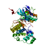 8efjC  8vmhC  8vmmC  8vs0C  8vt6C 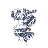 8vtfC  8vwmC C: citing same article ( |
|---|---|
| Similar structure data |
-Links
 PDBj
PDBj


- Assembly
| Deposited unit | 
| ||||||||||||
|---|---|---|---|---|---|---|---|---|---|---|---|---|---|
| 1 |
| ||||||||||||
| Unit cell |
|
-Components
| #1: Protein | Mass: 52649.332 Da / Num. of mol.: 1 Source method: isolated from a genetically manipulated source Source: (gene. exp.) Homo sapiens (human) / Gene: MAPK10, JNK3, JNK3A, PRKM10, SAPK1B / Production host:  References: UniProt: P53779, mitogen-activated protein kinase |
|---|---|
| #2: Chemical | ChemComp-WHE / ( Mass: 469.962 Da / Num. of mol.: 1 / Source method: obtained synthetically / Formula: C23H21ClFN5OS / Feature type: SUBJECT OF INVESTIGATION |
| #3: Water | ChemComp-HOH /  Mass: 18.015 Da / Num. of mol.: 178 / Source method: isolated from a natural source / Formula: H2O Mass: 18.015 Da / Num. of mol.: 178 / Source method: isolated from a natural source / Formula: H2O |
| Has ligand of interest | Y |
| Has protein modification | N |
-Experimental details
-Experiment
| Experiment | Method: X-RAY DIFFRACTION / Number of used crystals: 1 |
|---|
- Sample preparation
| Crystal | Density Matthews: 1.9 Å3/Da / Density meas: 35.38 Mg/m3 / Density % sol: 35.08 % |
|---|---|
| Crystal grow | Temperature: 277.15 K / Method: vapor diffusion / Details: 0.2 M ammonium tartrate pH 7.2, 18 % PEG 3350 |
-Data collection
| Diffraction | Mean temperature: 100 K / Serial crystal experiment: N |
|---|---|
| Diffraction source | Source: SYNCHROTRON / Site: SSRL / Beamline: BL12-2 / Wavelength: 1 Å |
| Detector | Type: DECTRIS PILATUS 6M / Detector: PIXEL / Date: Jan 19, 2020 |
| Radiation | Protocol: SINGLE WAVELENGTH / Monochromatic (M) / Laue (L): M / Scattering type: x-ray |
| Radiation wavelength | Wavelength: 1 Å / Relative weight: 1 |
| Reflection | Resolution: 2.06→59.27 Å / Num. obs: 25184 / % possible obs: 99.7 % / Redundancy: 4.1 % / Biso Wilson estimate: 28.81 Å2 / CC1/2: 0.99 / Net I/σ(I): 2.8 |
| Reflection shell | Resolution: 2.06→2.14 Å / Num. unique obs: 2480 / CC1/2: 0.77 |
- Processing
| Software |
| ||||||||||||||||||||||||||||||||||||||||||||||||||||||||||||||||||||||
|---|---|---|---|---|---|---|---|---|---|---|---|---|---|---|---|---|---|---|---|---|---|---|---|---|---|---|---|---|---|---|---|---|---|---|---|---|---|---|---|---|---|---|---|---|---|---|---|---|---|---|---|---|---|---|---|---|---|---|---|---|---|---|---|---|---|---|---|---|---|---|---|
| Refinement | Method to determine structure: MOLECULAR REPLACEMENT / Resolution: 2.07→53.77 Å / SU ML: 0.2587 / Cross valid method: FREE R-VALUE / σ(F): 1.34 / Phase error: 28.8128 Stereochemistry target values: GeoStd + Monomer Library + CDL v1.2
| ||||||||||||||||||||||||||||||||||||||||||||||||||||||||||||||||||||||
| Solvent computation | Shrinkage radii: 0.9 Å / VDW probe radii: 1.11 Å / Solvent model: FLAT BULK SOLVENT MODEL | ||||||||||||||||||||||||||||||||||||||||||||||||||||||||||||||||||||||
| Displacement parameters | Biso mean: 40.42 Å2 | ||||||||||||||||||||||||||||||||||||||||||||||||||||||||||||||||||||||
| Refinement step | Cycle: LAST / Resolution: 2.07→53.77 Å
| ||||||||||||||||||||||||||||||||||||||||||||||||||||||||||||||||||||||
| Refine LS restraints |
| ||||||||||||||||||||||||||||||||||||||||||||||||||||||||||||||||||||||
| LS refinement shell |
|
