 Movie
Movie Controller

[English] 日本語
 Yorodumi
- PDB-8vib: CW Flagellar Switch Complex - FliF, FliG, FliM, and FliN forming ... -
+ Open data

- Basic information
| Entry | Database: PDB / ID: 8vib | ||||||||||||||||||||||||||||||||||||||||||||||||
|---|---|---|---|---|---|---|---|---|---|---|---|---|---|---|---|---|---|---|---|---|---|---|---|---|---|---|---|---|---|---|---|---|---|---|---|---|---|---|---|---|---|---|---|---|---|---|---|---|---|
| Title | CW Flagellar Switch Complex - FliF, FliG, FliM, and FliN forming single subunit of the C-ring from Salmonella | ||||||||||||||||||||||||||||||||||||||||||||||||
 Components Components |
| ||||||||||||||||||||||||||||||||||||||||||||||||
 Keywords Keywords | MOTOR PROTEIN / Domain Swap / Symmetry mismatch / Flagellar component / Switch complex | ||||||||||||||||||||||||||||||||||||||||||||||||
| Function / homology |  Function and homology information Function and homology informationregulation of bacterial-type flagellum-dependent cell motility / bacterial-type flagellum basal body, MS ring / bacterial-type flagellum basal body / bacterial-type flagellum-dependent swarming motility / positive chemotaxis / cytoskeletal motor activity / bacterial-type flagellum-dependent cell motility / phosphoprotein phosphatase activity / chemotaxis / plasma membrane Similarity search - Function | ||||||||||||||||||||||||||||||||||||||||||||||||
| Biological species |  Salmonella enterica subsp. enterica serovar Typhimurium (bacteria) Salmonella enterica subsp. enterica serovar Typhimurium (bacteria) | ||||||||||||||||||||||||||||||||||||||||||||||||
| Method | ELECTRON MICROSCOPY / single particle reconstruction / cryo EM / Resolution: 4.6 Å | ||||||||||||||||||||||||||||||||||||||||||||||||
 Authors Authors | Singh, P.K. / Iverson, T.M. | ||||||||||||||||||||||||||||||||||||||||||||||||
| Funding support |  United States, 1items United States, 1items
| ||||||||||||||||||||||||||||||||||||||||||||||||
 Citation Citation | Journal: Nat Microbiol / Year: 2024 Title: CryoEM structures reveal how the bacterial flagellum rotates and switches direction. Authors: Prashant K Singh / Pankaj Sharma / Oshri Afanzar / Margo H Goldfarb / Elena Maklashina / Michael Eisenbach / Gary Cecchini / T M Iverson /  Abstract: Bacterial chemotaxis requires bidirectional flagellar rotation at different rates. Rotation is driven by a flagellar motor, which is a supercomplex containing multiple rings. Architectural ...Bacterial chemotaxis requires bidirectional flagellar rotation at different rates. Rotation is driven by a flagellar motor, which is a supercomplex containing multiple rings. Architectural uncertainty regarding the cytoplasmic C-ring, or 'switch', limits our understanding of how the motor transmits torque and direction to the flagellar rod. Here we report cryogenic electron microscopy structures for Salmonella enterica serovar typhimurium inner membrane MS-ring and C-ring in a counterclockwise pose (4.0 Å) and isolated C-ring in a clockwise pose alone (4.6 Å) and bound to a regulator (5.9 Å). Conformational differences between rotational poses include a 180° shift in FliF/FliG domains that rotates the outward-facing MotA/B binding site to inward facing. The regulator has specificity for the clockwise pose by bridging elements unique to this conformation. We used these structures to propose how the switch reverses rotation and transmits torque to the flagellum, which advances the understanding of bacterial chemotaxis and bidirectional motor rotation. | ||||||||||||||||||||||||||||||||||||||||||||||||
| History |
|
- Structure visualization
| Structure viewer | Molecule: MolmilJmol/JSmol |
|---|
- Downloads & links
-Download
| PDBx/mmCIF format | 8vib.cif.gz | 152.6 KB | Display | PDBx/mmCIF format |
|---|---|---|---|---|
| PDB format | pdb8vib.ent.gz | Display | PDB format | |
| PDBx/mmJSON format | 8vib.json.gz | Tree view | PDBx/mmJSON format | |
| Others |  Other downloads Other downloads |
-Validation report
| Arichive directory | https://data.pdbj.org/pub/pdb/validation_reports/vi/8vibftp://data.pdbj.org/pub/pdb/validation_reports/vi/8vib | HTTPS FTP |
|---|
-Related structure data
| Related structure data |  43256MC 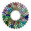 8t8pC 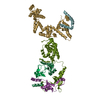 8vidC 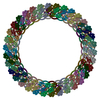 8vkqC 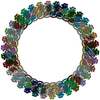 8vkrC M: map data used to model this data C: citing same article ( |
|---|---|
| Similar structure data |


-Links
 PDBj
PDBj
- Assembly
| Deposited unit | 
|
|---|---|
| 1 |
|
-Components
| #1: Protein | Mass: 61295.645 Da / Num. of mol.: 1 Source method: isolated from a genetically manipulated source Source: (gene. exp.) Salmonella enterica subsp. enterica serovar Typhimurium (bacteria)Gene: fliF, fla AII.1, fla BI, STM1969 / Production host: | ||
|---|---|---|---|
| #2: Protein | Mass: 36890.957 Da / Num. of mol.: 1 Source method: isolated from a genetically manipulated source Source: (gene. exp.) Salmonella enterica subsp. enterica serovar Typhimurium (bacteria)Gene: fliG, fla AII.2, fla BII, STM1970 / Production host: | ||
| #3: Protein | Mass: 37901.066 Da / Num. of mol.: 1 Source method: isolated from a genetically manipulated source Source: (gene. exp.) Salmonella enterica subsp. enterica serovar Typhimurium (bacteria)Gene: fliM, AAB27_20135, AKH62_22360, AU613_23675, AVC05_18485, B1P38_23905, CE70_09675, CFF59_05120, DD95_23260, DMO92_23265, DPF41_19480, DPS76_15080, DRM14_21765, DU071_20935, E0935_21110, EER35_ ...Gene: fliM, AAB27_20135, AKH62_22360, AU613_23675, AVC05_18485, B1P38_23905, CE70_09675, CFF59_05120, DD95_23260, DMO92_23265, DPF41_19480, DPS76_15080, DRM14_21765, DU071_20935, E0935_21110, EER35_17170, F3R12_13395, GB466_16335, SE14_02118 Production host: | ||
| #4: Protein | Mass: 14801.823 Da / Num. of mol.: 3 Source method: isolated from a genetically manipulated source Source: (gene. exp.) Salmonella enterica subsp. enterica serovar Typhimurium (bacteria)Gene: fliN, flaN, motD, STM1977 / Production host: Has protein modification | N | |
-Experimental details
-Experiment
| Experiment | Method: ELECTRON MICROSCOPY |
|---|---|
| EM experiment | Aggregation state: PARTICLE / 3D reconstruction method: single particle reconstruction |
- Sample preparation
| Component | Name: Flagellar C-ring single subunit containing FliF, FliG, FliM, and FliN Type: COMPLEX / Entity ID: all / Source: RECOMBINANT |
|---|---|
| Molecular weight | Value: 3.5 MDa / Experimental value: NO |
| Source (natural) | Organism: Salmonella enterica subsp. enterica serovar Typhimurium (bacteria) |
| Source (recombinant) | Organism: |
| Buffer solution | pH: 7.5 |
| Specimen | Embedding applied: NO / Shadowing applied: NO / Staining applied: NO / Vitrification applied: YES |
| Vitrification | Cryogen name: ETHANE |
- Electron microscopy imaging
| Experimental equipment |  Model: Titan Krios / Image courtesy: FEI Company |
|---|---|
| Microscopy | Model: TFS KRIOS |
| Electron gun | Electron source:  FIELD EMISSION GUN / Accelerating voltage: 300 kV / Illumination mode: FLOOD BEAM FIELD EMISSION GUN / Accelerating voltage: 300 kV / Illumination mode: FLOOD BEAM |
| Electron lens | Mode: BRIGHT FIELD / Nominal defocus max: 2000 nm / Nominal defocus min: 1000 nm |
| Image recording | Electron dose: 56.323 e/Å2 / Film or detector model: GATAN K3 (6k x 4k) |
- Processing
| EM software | Name: PHENIX / Version: 1.20.1_4487: / Category: model refinement |
|---|---|
| CTF correction | Type: PHASE FLIPPING AND AMPLITUDE CORRECTION |
| 3D reconstruction | Resolution: 4.6 Å / Resolution method: FSC 0.143 CUT-OFF / Num. of particles: 7201 / Symmetry type: POINT |
