 Movie
Movie Controller


+ Open data

- Basic information
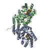
| Entry | Database: PDB / ID: 8vcw | ||||||
|---|---|---|---|---|---|---|---|
| Title | X-Ray Crystal Structure of the biotin synthase from B. obeum | ||||||
 Components Components | Biotin synthase | ||||||
 Keywords Keywords | TRANSFERASE / biotin / iron sulfur cluster / desthiobiotin / radical SAM | ||||||
| Function / homology |  Function and homology information Function and homology informationbiotin synthase / biotin synthase activity / biotin biosynthetic process / 2 iron, 2 sulfur cluster binding / 4 iron, 4 sulfur cluster binding / iron ion binding Similarity search - Function | ||||||
| Biological species |  Blautia obeum (bacteria) Blautia obeum (bacteria) | ||||||
| Method |  X-RAY DIFFRACTION / SYNCHROTRON / MOLECULAR REPLACEMENT / Resolution: 1.35 Å X-RAY DIFFRACTION / SYNCHROTRON / MOLECULAR REPLACEMENT / Resolution: 1.35 Å | ||||||
 Authors Authors | Lachowicz, J.C. / Grove, T.L. | ||||||
| Funding support |  United States, 1items United States, 1items
| ||||||
 Citation Citation | Journal: J.Am.Chem.Soc. / Year: 2024 Title: Discovery of a Biotin Synthase That Utilizes an Auxiliary 4Fe-5S Cluster for Sulfur Insertion. Authors: Lachowicz, J.C. / Lennox-Hvenekilde, D. / Myling-Petersen, N. / Salomonsen, B. / Verkleij, G. / Acevedo-Rocha, C.G. / Caddell, B. / Gronenberg, L.S. / Almo, S.C. / Sommer, M.O.A. / Genee, H.J. / Grove, T.L. | ||||||
| History |
|
- Structure visualization
| Structure viewer | Molecule: MolmilJmol/JSmol |
|---|
- Downloads & links
-Download
| PDBx/mmCIF format | 8vcw.cif.gz | 94.9 KB | Display | PDBx/mmCIF format |
|---|---|---|---|---|
| PDB format | pdb8vcw.ent.gz | 67.6 KB | Display | PDB format |
| PDBx/mmJSON format | 8vcw.json.gz | Tree view | PDBx/mmJSON format | |
| Others |  Other downloads Other downloads |
-Validation report
| Arichive directory | https://data.pdbj.org/pub/pdb/validation_reports/vc/8vcwftp://data.pdbj.org/pub/pdb/validation_reports/vc/8vcw | HTTPS FTP |
|---|
-Related structure data
-Links
 PDBj
PDBj
- Assembly
| Deposited unit | 
| ||||||||
|---|---|---|---|---|---|---|---|---|---|
| 1 |
| ||||||||
| Unit cell |
|
-Components
-Protein , 1 types, 1 molecules A
| #1: Protein | Mass: 36331.398 Da / Num. of mol.: 1 Source method: isolated from a genetically manipulated source Details: Contains a 4 iron:5 sulfur cluster / Source: (gene. exp.) Blautia obeum (bacteria) / Gene: bioB, RUMOBE_02699 / Plasmid: pSGCHis / Production host: |
|---|
-Non-polymers , 8 types, 426 molecules 


| #2: Chemical | ChemComp-SAM /  Mass: 398.437 Da / Num. of mol.: 1 / Source method: obtained synthetically / Formula: C15H22N6O5S / Feature type: SUBJECT OF INVESTIGATION Mass: 398.437 Da / Num. of mol.: 1 / Source method: obtained synthetically / Formula: C15H22N6O5S / Feature type: SUBJECT OF INVESTIGATION |
|---|---|
| #3: Chemical | ChemComp-BTB /  Mass: 209.240 Da / Num. of mol.: 1 / Source method: obtained synthetically / Formula: C8H19NO5 / Comment: pH buffer*YM Mass: 209.240 Da / Num. of mol.: 1 / Source method: obtained synthetically / Formula: C8H19NO5 / Comment: pH buffer*YM |
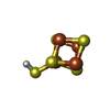
| #4: Chemical | ChemComp-Q46 /  Mass: 384.713 Da / Num. of mol.: 1 / Source method: obtained synthetically / Formula: Fe4HS5 / Feature type: SUBJECT OF INVESTIGATION Mass: 384.713 Da / Num. of mol.: 1 / Source method: obtained synthetically / Formula: Fe4HS5 / Feature type: SUBJECT OF INVESTIGATION |
| #5: Chemical | ChemComp-SF4 /  Mass: 351.640 Da / Num. of mol.: 1 / Source method: obtained synthetically / Formula: Fe4S4 / Feature type: SUBJECT OF INVESTIGATION Mass: 351.640 Da / Num. of mol.: 1 / Source method: obtained synthetically / Formula: Fe4S4 / Feature type: SUBJECT OF INVESTIGATION |
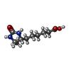
| #6: Chemical | ChemComp-DTB /  Mass: 214.262 Da / Num. of mol.: 1 / Source method: obtained synthetically / Formula: C10H18N2O3 / Feature type: SUBJECT OF INVESTIGATION Mass: 214.262 Da / Num. of mol.: 1 / Source method: obtained synthetically / Formula: C10H18N2O3 / Feature type: SUBJECT OF INVESTIGATION |
| #7: Chemical | ChemComp-FE /  Mass: 55.845 Da / Num. of mol.: 1 / Source method: obtained synthetically / Formula: Fe Mass: 55.845 Da / Num. of mol.: 1 / Source method: obtained synthetically / Formula: Fe |
| #8: Chemical | ChemComp-CL /  Mass: 35.453 Da / Num. of mol.: 1 / Source method: obtained synthetically / Formula: Cl Mass: 35.453 Da / Num. of mol.: 1 / Source method: obtained synthetically / Formula: Cl |
| #9: Water | ChemComp-HOH / Mass: 18.015 Da / Num. of mol.: 419 / Source method: isolated from a natural source / Formula: H2O |
-Details
| Has ligand of interest | Y |
|---|---|
| Has protein modification | N |
-Experimental details
-Experiment
| Experiment | Method: X-RAY DIFFRACTION / Number of used crystals: 1 |
|---|
- Sample preparation
| Crystal | Density Matthews: 2.16 Å3/Da / Density % sol: 43.08 % |
|---|---|
| Crystal grow | Temperature: 298 K / Method: vapor diffusion, sitting drop / pH: 5.5 Details: 0.4uL protein plus 0.4 uL 100mM Bis-Tri pH 5.5, 25% PEG 3350, 0.2M NaCl equilibrated against a well of 0.5M LiCl |
-Data collection
| Diffraction | Mean temperature: 100 K / Serial crystal experiment: N |
|---|---|
| Diffraction source | Source: SYNCHROTRON / Site: NSLS-II / Beamline: 17-ID-2 / Wavelength: 0.9793 Å |
| Detector | Type: DECTRIS EIGER X 16M / Detector: PIXEL / Date: Oct 7, 2022 / Details: mirrors |
| Radiation | Monochromator: Si (111) / Protocol: SINGLE WAVELENGTH / Monochromatic (M) / Laue (L): M / Scattering type: x-ray |
| Radiation wavelength | Wavelength: 0.9793 Å / Relative weight: 1 |
| Reflection | Resolution: 1.35→80.94 Å / Num. obs: 67982 / % possible obs: 96.9 % / Redundancy: 4.1 % / Biso Wilson estimate: 11.8 Å2 / CC1/2: 0.998 / Rmerge(I) obs: 0.067 / Rpim(I) all: 0.036 / Rrim(I) all: 0.076 / Χ2: 1.05 / Net I/σ(I): 12 |
| Reflection shell | Resolution: 1.35→1.37 Å / Redundancy: 2.6 % / Rmerge(I) obs: 0.436 / Mean I/σ(I) obs: 2.4 / Num. unique obs: 2523 / CC1/2: 0.741 / Rpim(I) all: 0.304 / Rrim(I) all: 0.535 / Χ2: 1.17 / % possible all: 75 |
- Processing
| Software |
| ||||||||||||||||||||||||||||||||||||||||||||||||||||||||||||||||||||||||||||||||||||||||||||||||||||||||||||||||||||||||||||||||||||||||||||||||||||||||||||||||||||||||
|---|---|---|---|---|---|---|---|---|---|---|---|---|---|---|---|---|---|---|---|---|---|---|---|---|---|---|---|---|---|---|---|---|---|---|---|---|---|---|---|---|---|---|---|---|---|---|---|---|---|---|---|---|---|---|---|---|---|---|---|---|---|---|---|---|---|---|---|---|---|---|---|---|---|---|---|---|---|---|---|---|---|---|---|---|---|---|---|---|---|---|---|---|---|---|---|---|---|---|---|---|---|---|---|---|---|---|---|---|---|---|---|---|---|---|---|---|---|---|---|---|---|---|---|---|---|---|---|---|---|---|---|---|---|---|---|---|---|---|---|---|---|---|---|---|---|---|---|---|---|---|---|---|---|---|---|---|---|---|---|---|---|---|---|---|---|---|---|---|---|
| Refinement | Method to determine structure: MOLECULAR REPLACEMENT / Resolution: 1.35→48.45 Å / SU ML: 0.12 / Cross valid method: FREE R-VALUE / σ(F): 1.33 / Phase error: 16.59 / Stereochemistry target values: ML
| ||||||||||||||||||||||||||||||||||||||||||||||||||||||||||||||||||||||||||||||||||||||||||||||||||||||||||||||||||||||||||||||||||||||||||||||||||||||||||||||||||||||||
| Solvent computation | Shrinkage radii: 0.9 Å / VDW probe radii: 1.1 Å / Solvent model: FLAT BULK SOLVENT MODEL | ||||||||||||||||||||||||||||||||||||||||||||||||||||||||||||||||||||||||||||||||||||||||||||||||||||||||||||||||||||||||||||||||||||||||||||||||||||||||||||||||||||||||
| Displacement parameters | Biso mean: 12.1 Å2 | ||||||||||||||||||||||||||||||||||||||||||||||||||||||||||||||||||||||||||||||||||||||||||||||||||||||||||||||||||||||||||||||||||||||||||||||||||||||||||||||||||||||||
| Refinement step | Cycle: LAST / Resolution: 1.35→48.45 Å
| ||||||||||||||||||||||||||||||||||||||||||||||||||||||||||||||||||||||||||||||||||||||||||||||||||||||||||||||||||||||||||||||||||||||||||||||||||||||||||||||||||||||||
| Refine LS restraints |
| ||||||||||||||||||||||||||||||||||||||||||||||||||||||||||||||||||||||||||||||||||||||||||||||||||||||||||||||||||||||||||||||||||||||||||||||||||||||||||||||||||||||||
| LS refinement shell |
|
