 Movie
Movie Controller


+ Open data

- Basic information
| Entry | Database: PDB / ID: 8vdw | ||||||
|---|---|---|---|---|---|---|---|
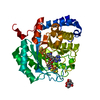
| Title | X-Ray Crystal Structure of the biotin synthase from V. parvula | ||||||
 Components Components | Biotin synthase | ||||||
 Keywords Keywords | TRANSFERASE / radical SAM / iron-sulfur cluster / sulfur insertion / 4 iron-5 sulfur cluster | ||||||
| Function / homology |  Function and homology information Function and homology informationbiotin synthase / biotin synthase activity / biotin biosynthetic process / 2 iron, 2 sulfur cluster binding / 4 iron, 4 sulfur cluster binding / iron ion binding Similarity search - Function | ||||||
| Biological species |  Veillonella parvula (bacteria) Veillonella parvula (bacteria) | ||||||
| Method |  X-RAY DIFFRACTION / SYNCHROTRON / MOLECULAR REPLACEMENT / Resolution: 1.807 Å X-RAY DIFFRACTION / SYNCHROTRON / MOLECULAR REPLACEMENT / Resolution: 1.807 Å | ||||||
 Authors Authors | Lachowicz, J.C. / Grove, T.L. | ||||||
| Funding support |  United States, 1items United States, 1items
| ||||||
 Citation Citation | Journal: J.Am.Chem.Soc. / Year: 2024 Title: Discovery of a Biotin Synthase That Utilizes an Auxiliary 4Fe-5S Cluster for Sulfur Insertion. Authors: Lachowicz, J.C. / Lennox-Hvenekilde, D. / Myling-Petersen, N. / Salomonsen, B. / Verkleij, G. / Acevedo-Rocha, C.G. / Caddell, B. / Gronenberg, L.S. / Almo, S.C. / Sommer, M.O.A. / Genee, H.J. / Grove, T.L. | ||||||
| History |
|
- Structure visualization
| Structure viewer | Molecule: MolmilJmol/JSmol |
|---|
- Downloads & links
-Download
| PDBx/mmCIF format | 8vdw.cif.gz | 161.7 KB | Display | PDBx/mmCIF format |
|---|---|---|---|---|
| PDB format | pdb8vdw.ent.gz | 123.3 KB | Display | PDB format |
| PDBx/mmJSON format | 8vdw.json.gz | Tree view | PDBx/mmJSON format | |
| Others |  Other downloads Other downloads |
-Validation report
| Arichive directory | https://data.pdbj.org/pub/pdb/validation_reports/vd/8vdwftp://data.pdbj.org/pub/pdb/validation_reports/vd/8vdw | HTTPS FTP |
|---|
-Related structure data
-Links
 PDBj
PDBj
- Assembly
| Deposited unit | 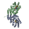
| ||||||||
|---|---|---|---|---|---|---|---|---|---|
| 1 |
| ||||||||
| Unit cell |
|
-Components
-Protein , 1 types, 2 molecules AB
| #1: Protein | Mass: 40487.883 Da / Num. of mol.: 2 Source method: isolated from a genetically manipulated source Source: (gene. exp.) Veillonella parvula (bacteria) / Gene: bioB, HSIVP1_982 / Plasmid: psGCHis / Production host: |
|---|
-Non-polymers , 6 types, 564 molecules 


| #2: Chemical |  Mass: 384.713 Da / Num. of mol.: 2 / Source method: obtained synthetically / Formula: Fe4HS5 / Feature type: SUBJECT OF INVESTIGATION Mass: 384.713 Da / Num. of mol.: 2 / Source method: obtained synthetically / Formula: Fe4HS5 / Feature type: SUBJECT OF INVESTIGATION#3: Chemical |  Mass: 398.437 Da / Num. of mol.: 2 / Source method: obtained synthetically / Formula: C15H22N6O5S / Feature type: SUBJECT OF INVESTIGATION Mass: 398.437 Da / Num. of mol.: 2 / Source method: obtained synthetically / Formula: C15H22N6O5S / Feature type: SUBJECT OF INVESTIGATION#4: Chemical |  Mass: 351.640 Da / Num. of mol.: 2 / Source method: obtained synthetically / Formula: Fe4S4 / Feature type: SUBJECT OF INVESTIGATION Mass: 351.640 Da / Num. of mol.: 2 / Source method: obtained synthetically / Formula: Fe4S4 / Feature type: SUBJECT OF INVESTIGATION#5: Chemical |  Mass: 214.262 Da / Num. of mol.: 2 / Source method: obtained synthetically / Formula: C10H18N2O3 / Feature type: SUBJECT OF INVESTIGATION Mass: 214.262 Da / Num. of mol.: 2 / Source method: obtained synthetically / Formula: C10H18N2O3 / Feature type: SUBJECT OF INVESTIGATION#6: Chemical | ChemComp-NA / |  Mass: 22.990 Da / Num. of mol.: 1 / Source method: obtained synthetically / Formula: Na Mass: 22.990 Da / Num. of mol.: 1 / Source method: obtained synthetically / Formula: Na#7: Water | ChemComp-HOH / | Mass: 18.015 Da / Num. of mol.: 555 / Source method: isolated from a natural source / Formula: H2O |
|---|
-Details
| Has ligand of interest | Y |
|---|
-Experimental details
-Experiment
| Experiment | Method: X-RAY DIFFRACTION / Number of used crystals: 1 |
|---|
- Sample preparation
| Crystal | Density Matthews: 2.15 Å3/Da / Density % sol: 42.82 % |
|---|---|
| Crystal grow | Temperature: 298 K / Method: vapor diffusion, sitting drop / pH: 5.5 / Details: 0.1M Bis-Tris pH 5.5, 0.2M NH4OAc, 25% PEG 3350 |
-Data collection
| Diffraction | Mean temperature: 100 K / Serial crystal experiment: N |
|---|---|
| Diffraction source | Source: SYNCHROTRON / Site: NSLS-II / Beamline: 17-ID-1 / Wavelength: 0.9793 Å |
| Detector | Type: DECTRIS EIGER X 16M / Detector: PIXEL / Date: Dec 11, 2018 / Details: mirrors |
| Radiation | Monochromator: Si (111) / Protocol: SINGLE WAVELENGTH / Monochromatic (M) / Laue (L): M / Scattering type: x-ray |
| Radiation wavelength | Wavelength: 0.9793 Å / Relative weight: 1 |
| Reflection | Resolution: 1.807→29.6 Å / Num. obs: 60902 / % possible obs: 99.7 % / Redundancy: 6.7 % / CC1/2: 0.999 / Rmerge(I) obs: 0.094 / Rpim(I) all: 0.039 / Rrim(I) all: 0.102 / Net I/σ(I): 12.6 |
| Reflection shell | Resolution: 1.81→1.85 Å / % possible obs: 96 % / Redundancy: 6.1 % / Rmerge(I) obs: 0.757 / Num. measured all: 26113 / Num. unique obs: 4262 / CC1/2: 0.855 / Rpim(I) all: 0.328 / Rrim(I) all: 0.827 / Net I/σ(I) obs: 2 |
- Processing
| Software |
| ||||||||||||||||||||||||||||||||||||||||||||||||||||||||||||||||||||||||||||||||||||||||||||||||||||||||||||||||||||||||||||||||||||||||||||||||||||||||||||||||||||||||||||||||||||||
|---|---|---|---|---|---|---|---|---|---|---|---|---|---|---|---|---|---|---|---|---|---|---|---|---|---|---|---|---|---|---|---|---|---|---|---|---|---|---|---|---|---|---|---|---|---|---|---|---|---|---|---|---|---|---|---|---|---|---|---|---|---|---|---|---|---|---|---|---|---|---|---|---|---|---|---|---|---|---|---|---|---|---|---|---|---|---|---|---|---|---|---|---|---|---|---|---|---|---|---|---|---|---|---|---|---|---|---|---|---|---|---|---|---|---|---|---|---|---|---|---|---|---|---|---|---|---|---|---|---|---|---|---|---|---|---|---|---|---|---|---|---|---|---|---|---|---|---|---|---|---|---|---|---|---|---|---|---|---|---|---|---|---|---|---|---|---|---|---|---|---|---|---|---|---|---|---|---|---|---|---|---|---|---|
| Refinement | Method to determine structure: MOLECULAR REPLACEMENT / Resolution: 1.807→29.6 Å / Cor.coef. Fo:Fc: 0.966 / Cor.coef. Fo:Fc free: 0.942 / SU B: 3.914 / SU ML: 0.112 / Cross valid method: THROUGHOUT / ESU R: 0.142 / ESU R Free: 0.137 / Stereochemistry target values: MAXIMUM LIKELIHOOD / Details: HYDROGENS HAVE BEEN ADDED IN THE RIDING POSITIONS
| ||||||||||||||||||||||||||||||||||||||||||||||||||||||||||||||||||||||||||||||||||||||||||||||||||||||||||||||||||||||||||||||||||||||||||||||||||||||||||||||||||||||||||||||||||||||
| Solvent computation | Ion probe radii: 0.8 Å / Shrinkage radii: 0.8 Å / VDW probe radii: 1.2 Å / Solvent model: MASK | ||||||||||||||||||||||||||||||||||||||||||||||||||||||||||||||||||||||||||||||||||||||||||||||||||||||||||||||||||||||||||||||||||||||||||||||||||||||||||||||||||||||||||||||||||||||
| Displacement parameters | Biso mean: 27.7 Å2
| ||||||||||||||||||||||||||||||||||||||||||||||||||||||||||||||||||||||||||||||||||||||||||||||||||||||||||||||||||||||||||||||||||||||||||||||||||||||||||||||||||||||||||||||||||||||
| Refinement step | Cycle: 1 / Resolution: 1.807→29.6 Å
| ||||||||||||||||||||||||||||||||||||||||||||||||||||||||||||||||||||||||||||||||||||||||||||||||||||||||||||||||||||||||||||||||||||||||||||||||||||||||||||||||||||||||||||||||||||||
| Refine LS restraints |
|