 Movie
Movie Controller


+ Open data

- Basic information
| Entry | Database: PDB / ID: 8v3o | ||||||
|---|---|---|---|---|---|---|---|
| Title | CCP5 in complex with Glu-P-peptide 1 transition state analog | ||||||
 Components Components |
| ||||||
 Keywords Keywords | HYDROLASE/INHIBITOR / carboxypeptidase deglutamylation branch glutamate removal microtubule / HYDROLASE / HYDROLASE-INHIBITOR complex | ||||||
| Function / homology |  Function and homology information Function and homology informationtubulin-glutamate carboxypeptidase / protein deglutamylation / C-terminal protein deglutamylation / protein side chain deglutamylation / protein branching point deglutamylation / Post-chaperonin tubulin folding pathway / Cilium Assembly / Carboxyterminal post-translational modifications of tubulin / Microtubule-dependent trafficking of connexons from Golgi to the plasma membrane / Sealing of the nuclear envelope (NE) by ESCRT-III ...tubulin-glutamate carboxypeptidase / protein deglutamylation / C-terminal protein deglutamylation / protein side chain deglutamylation / protein branching point deglutamylation / Post-chaperonin tubulin folding pathway / Cilium Assembly / Carboxyterminal post-translational modifications of tubulin / Microtubule-dependent trafficking of connexons from Golgi to the plasma membrane / Sealing of the nuclear envelope (NE) by ESCRT-III / Intraflagellar transport / Formation of tubulin folding intermediates by CCT/TriC / Gap junction assembly / Kinesins / Prefoldin mediated transfer of substrate to CCT/TriC / Assembly and cell surface presentation of NMDA receptors / COPI-independent Golgi-to-ER retrograde traffic / COPI-dependent Golgi-to-ER retrograde traffic / Recycling pathway of L1 / Hydrolases; Acting on peptide bonds (peptidases); Metallocarboxypeptidases / RHO GTPases activate IQGAPs / Hedgehog 'off' state / intercellular bridge / COPI-mediated anterograde transport / Activation of AMPK downstream of NMDARs / metallocarboxypeptidase activity / MHC class II antigen presentation / Recruitment of NuMA to mitotic centrosomes / Mitotic Prometaphase / HSP90 chaperone cycle for steroid hormone receptors (SHR) in the presence of ligand / EML4 and NUDC in mitotic spindle formation / Resolution of Sister Chromatid Cohesion / Translocation of SLC2A4 (GLUT4) to the plasma membrane / tubulin binding / RHO GTPases Activate Formins / cerebral cortex development / PKR-mediated signaling / structural constituent of cytoskeleton / microtubule cytoskeleton organization / neuron migration / HCMV Early Events / Aggrephagy / The role of GTSE1 in G2/M progression after G2 checkpoint / mitotic spindle / Separation of Sister Chromatids / mitotic cell cycle / extracellular vesicle / microtubule cytoskeleton / midbody / defense response to virus / microtubule / GTPase activity / GTP binding / proteolysis / extracellular exosome / zinc ion binding / metal ion binding / nucleus / cytoplasm / cytosol Similarity search - Function | ||||||
| Biological species |  Homo sapiens (human) Homo sapiens (human) | ||||||
| Method |  X-RAY DIFFRACTION / SYNCHROTRON / MOLECULAR REPLACEMENT / Resolution: 2.3 Å X-RAY DIFFRACTION / SYNCHROTRON / MOLECULAR REPLACEMENT / Resolution: 2.3 Å | ||||||
 Authors Authors | Chen, J. / Zehr, E.A. / Gruschus, J.M. / Szyk, A. / Liu, Y. / Tanner, M.E. / Tjandra, N. / Roll-Mecak, A. | ||||||
| Funding support |  United States, 1items United States, 1items
| ||||||
 Citation Citation | Journal: Nature / Year: 2024 Title: Tubulin code eraser CCP5 binds branch glutamates by substrate deformation. Authors: Jiayi Chen / Elena A Zehr / James M Gruschus / Agnieszka Szyk / Yanjie Liu / Martin E Tanner / Nico Tjandra / Antonina Roll-Mecak /  Abstract: Microtubule function is modulated by the tubulin code, diverse posttranslational modifications that are altered dynamically by writer and eraser enzymes. Glutamylation-the addition of branched ...Microtubule function is modulated by the tubulin code, diverse posttranslational modifications that are altered dynamically by writer and eraser enzymes. Glutamylation-the addition of branched (isopeptide-linked) glutamate chains-is the most evolutionarily widespread tubulin modification. It is introduced by tubulin tyrosine ligase-like enzymes and erased by carboxypeptidases of the cytosolic carboxypeptidase (CCP) family. Glutamylation homeostasis, achieved through the balance of writers and erasers, is critical for normal cell function, and mutations in CCPs lead to human disease. Here we report cryo-electron microscopy structures of the glutamylation eraser CCP5 in complex with the microtubule, and X-ray structures in complex with transition-state analogues. Combined with NMR analysis, these analyses show that CCP5 deforms the tubulin main chain into a unique turn that enables lock-and-key recognition of the branch glutamate in a cationic pocket that is unique to CCP family proteins. CCP5 binding of the sequences flanking the branch point primarily through peptide backbone atoms enables processing of diverse tubulin isotypes and non-tubulin substrates. Unexpectedly, CCP5 exhibits inefficient processing of an abundant β-tubulin isotype in the brain. This work provides an atomistic view into glutamate branch recognition and resolution, and sheds light on homeostasis of the tubulin glutamylation syntax. | ||||||
| History |
|
- Structure visualization
| Structure viewer | Molecule: MolmilJmol/JSmol |
|---|
- Downloads & links
-Download
| PDBx/mmCIF format | 8v3o.cif.gz | 116.4 KB | Display | PDBx/mmCIF format |
|---|---|---|---|---|
| PDB format | pdb8v3o.ent.gz | 85.9 KB | Display | PDB format |
| PDBx/mmJSON format | 8v3o.json.gz | Tree view | PDBx/mmJSON format | |
| Others |  Other downloads Other downloads |
-Validation report
| Arichive directory | https://data.pdbj.org/pub/pdb/validation_reports/v3/8v3oftp://data.pdbj.org/pub/pdb/validation_reports/v3/8v3o | HTTPS FTP |
|---|
-Related structure data
| Related structure data |  8v3mC  8v3nC  8v3pC  8v3qC  8v3rC  8v3sC  8v4kC 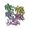 8v4lC 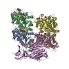 8v4mC C: citing same article ( |
|---|---|
| Similar structure data |


-Links
 PDBj
PDBj


- Assembly
| Deposited unit | 
| ||||||||
|---|---|---|---|---|---|---|---|---|---|
| 1 |
| ||||||||
| Unit cell |
|
-Components
-Protein / Protein/peptide , 2 types, 2 molecules AI
| #1: Protein | Mass: 59170.668 Da / Num. of mol.: 1 / Mutation: E516A,residues 339-424 replaced with a SGSGG loop Source method: isolated from a genetically manipulated source Source: (gene. exp.) Homo sapiens (human) / Gene: AGBL5, CCP5 / Plasmid: pFastBac / Details (production host): His_MBP_Asn10_TEV / Cell line (production host): Sf9 / Production host:   Spodoptera frugiperda (fall armyworm) / Strain (production host): IPLB-Sf-21-AE Spodoptera frugiperda (fall armyworm) / Strain (production host): IPLB-Sf-21-AEReferences: UniProt: Q8NDL9, Hydrolases; Acting on peptide bonds (peptidases); Metallocarboxypeptidases, tubulin-glutamate carboxypeptidase |
|---|---|
| #2: Protein/peptide | Mass: 1905.682 Da / Num. of mol.: 1 / Mutation: One of the glutamates replaced with BIX / Source method: obtained synthetically / Source: (synth.) Homo sapiens (human) / References: UniProt: Q13885 |
-Non-polymers , 4 types, 143 molecules 


| #3: Chemical | ChemComp-ZN /  Mass: 65.409 Da / Num. of mol.: 1 / Source method: obtained synthetically / Formula: Zn Mass: 65.409 Da / Num. of mol.: 1 / Source method: obtained synthetically / Formula: Zn | ||||
|---|---|---|---|---|---|
| #4: Chemical |  Mass: 134.087 Da / Num. of mol.: 2 / Source method: obtained synthetically / Formula: C4H6O5 Mass: 134.087 Da / Num. of mol.: 2 / Source method: obtained synthetically / Formula: C4H6O5#5: Chemical | ChemComp-K / |  Mass: 39.098 Da / Num. of mol.: 1 / Source method: obtained synthetically / Formula: K Mass: 39.098 Da / Num. of mol.: 1 / Source method: obtained synthetically / Formula: K#6: Water | ChemComp-HOH / | Mass: 18.015 Da / Num. of mol.: 139 / Source method: isolated from a natural source / Formula: H2O |
-Details
| Has ligand of interest | Y |
|---|---|
| Has protein modification | Y |
-Experimental details
-Experiment
| Experiment | Method: X-RAY DIFFRACTION / Number of used crystals: 1 |
|---|
- Sample preparation
| Crystal | Density Matthews: 2.26 Å3/Da / Density % sol: 45.49 % |
|---|---|
| Crystal grow | Temperature: 294 K / Method: vapor diffusion, hanging drop Details: 0.15 M DL-Malic acid, pH7.0, 0.1 M imidazole, pH7.0, 16% PEG MME 550 |
-Data collection
| Diffraction | Mean temperature: 93 K / Serial crystal experiment: N |
|---|---|
| Diffraction source | Source: SYNCHROTRON / Site: ALS / Beamline: 8.2.1 / Wavelength: 1 Å |
| Detector | Type: DECTRIS EIGER X 9M / Detector: PIXEL / Date: May 17, 2022 |
| Radiation | Protocol: SINGLE WAVELENGTH / Monochromatic (M) / Laue (L): M / Scattering type: x-ray |
| Radiation wavelength | Wavelength: 1 Å / Relative weight: 1 |
| Reflection | Resolution: 2.3→34.55 Å / Num. obs: 25384 / % possible obs: 100 % / Redundancy: 14.3 % / CC1/2: 0.998 / Rmerge(I) obs: 0.175 / Rrim(I) all: 0.187 / Net I/σ(I): 13.8 |
| Reflection shell | Resolution: 2.3→2.38 Å / Redundancy: 14.7 % / Rmerge(I) obs: 1.455 / Mean I/σ(I) obs: 2 / Num. unique obs: 2454 / CC1/2: 0.712 / Rrim(I) all: 1.56 / % possible all: 100 |
- Processing
| Software |
| |||||||||||||||||||||||||||||||||||||||||||||||||||||||||||||||
|---|---|---|---|---|---|---|---|---|---|---|---|---|---|---|---|---|---|---|---|---|---|---|---|---|---|---|---|---|---|---|---|---|---|---|---|---|---|---|---|---|---|---|---|---|---|---|---|---|---|---|---|---|---|---|---|---|---|---|---|---|---|---|---|---|
| Refinement | Method to determine structure: MOLECULAR REPLACEMENT / Resolution: 2.3→34.55 Å / SU ML: 0.21 / Cross valid method: FREE R-VALUE / σ(F): 1.34 / Phase error: 23.91 / Stereochemistry target values: ML
| |||||||||||||||||||||||||||||||||||||||||||||||||||||||||||||||
| Solvent computation | Shrinkage radii: 0.9 Å / VDW probe radii: 1.1 Å / Solvent model: FLAT BULK SOLVENT MODEL | |||||||||||||||||||||||||||||||||||||||||||||||||||||||||||||||
| Refinement step | Cycle: LAST / Resolution: 2.3→34.55 Å
| |||||||||||||||||||||||||||||||||||||||||||||||||||||||||||||||
| Refine LS restraints |
| |||||||||||||||||||||||||||||||||||||||||||||||||||||||||||||||
| LS refinement shell |
|
