Movie
Movie Controller

[English] 日本語
 Yorodumi
- EMDB-42948: Microtubule protofilament reconstruction in CCP5:microtubule clas... -
+ Open data

- Basic information
| Entry |  | |||||||||
|---|---|---|---|---|---|---|---|---|---|---|
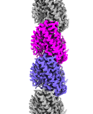
| Title | Microtubule protofilament reconstruction in CCP5:microtubule class#1 complex | |||||||||
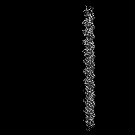
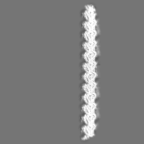
 Map data Map data | Protofilament reconstruction in class 1, b-factor sharpened | |||||||||
 Sample Sample |
| |||||||||
 Keywords Keywords | microtubules / cytoskeleton / STRUCTURAL PROTEIN | |||||||||
| Biological species |  | |||||||||
| Method | single particle reconstruction / cryo EM / Resolution: 3.0 Å | |||||||||
 Authors Authors | Chen J / Zehr EA / Gruschus JM / Szyk A / Liu Y / Tanner ME / Tjandra N / Roll-Mecak A | |||||||||
| Funding support |  United States, 1 items United States, 1 items
| |||||||||
 Citation Citation | Journal: Nature / Year: 2024 Title: Tubulin code eraser CCP5 binds branch glutamates by substrate deformation. Authors: Jiayi Chen / Elena A Zehr / James M Gruschus / Agnieszka Szyk / Yanjie Liu / Martin E Tanner / Nico Tjandra / Antonina Roll-Mecak /  Abstract: Microtubule function is modulated by the tubulin code, diverse posttranslational modifications that are altered dynamically by writer and eraser enzymes. Glutamylation-the addition of branched ...Microtubule function is modulated by the tubulin code, diverse posttranslational modifications that are altered dynamically by writer and eraser enzymes. Glutamylation-the addition of branched (isopeptide-linked) glutamate chains-is the most evolutionarily widespread tubulin modification. It is introduced by tubulin tyrosine ligase-like enzymes and erased by carboxypeptidases of the cytosolic carboxypeptidase (CCP) family. Glutamylation homeostasis, achieved through the balance of writers and erasers, is critical for normal cell function, and mutations in CCPs lead to human disease. Here we report cryo-electron microscopy structures of the glutamylation eraser CCP5 in complex with the microtubule, and X-ray structures in complex with transition-state analogues. Combined with NMR analysis, these analyses show that CCP5 deforms the tubulin main chain into a unique turn that enables lock-and-key recognition of the branch glutamate in a cationic pocket that is unique to CCP family proteins. CCP5 binding of the sequences flanking the branch point primarily through peptide backbone atoms enables processing of diverse tubulin isotypes and non-tubulin substrates. Unexpectedly, CCP5 exhibits inefficient processing of an abundant β-tubulin isotype in the brain. This work provides an atomistic view into glutamate branch recognition and resolution, and sheds light on homeostasis of the tubulin glutamylation syntax. | |||||||||
| History |
|
- Structure visualization
| Supplemental images |
|---|
- Downloads & links
-EMDB archive
| Map data | emd_42948.map.gz | 16.4 MB |  EMDB map data format EMDB map data format | |
|---|---|---|---|---|
| Header (meta data) | emd-42948-v30.xmlemd-42948.xml | 20.5 KB 20.5 KB | Display Display | EMDB header |
| FSC (resolution estimation) | emd_42948_fsc.xml | 21 KB | Display | FSC data file |
| Images |  emd_42948.png emd_42948.png | 93.1 KB | ||
| Masks | emd_42948_msk_1.map | 381.1 MB | Mask map | |
| Filedesc metadata | emd-42948.cif.gz | 4.8 KB | ||
| Others | emd_42948_additional_1.map.gzemd_42948_additional_2.map.gzemd_42948_half_map_1.map.gzemd_42948_half_map_2.map.gz | 299.7 MB 334.3 MB 301.9 MB 301.9 MB | ||
| Archive directory |  http://ftp.pdbj.org/pub/emdb/structures/EMD-42948ftp://ftp.pdbj.org/pub/emdb/structures/EMD-42948 http://ftp.pdbj.org/pub/emdb/structures/EMD-42948ftp://ftp.pdbj.org/pub/emdb/structures/EMD-42948 | HTTPS FTP |
-Related structure data
| Related structure data |  8v3mC  8v3nC  8v3oC 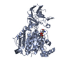 8v3pC 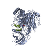 8v3qC 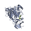 8v3rC 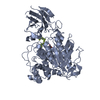 8v3sC  8v4kC 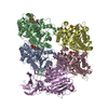 8v4lC 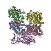 8v4mC C: citing same article ( |
|---|


-Links
| EMDB pages | EMDB (EBI/PDBe) / EMDataResource |
|---|
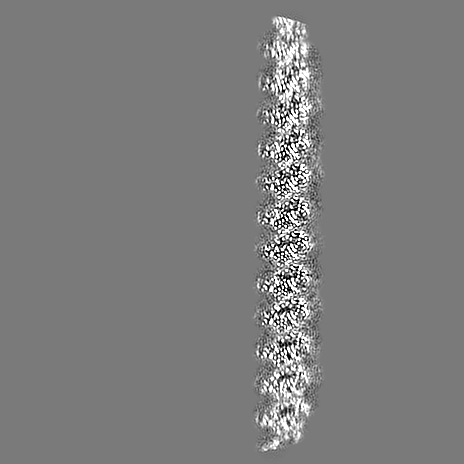
-Map
| File | Download / File: emd_42948.map.gz / Format: CCP4 / Size: 381.1 MB / Type: IMAGE STORED AS FLOATING POINT NUMBER (4 BYTES) | ||||||||||||||||||||||||||||||||||||
|---|---|---|---|---|---|---|---|---|---|---|---|---|---|---|---|---|---|---|---|---|---|---|---|---|---|---|---|---|---|---|---|---|---|---|---|---|---|
| Annotation | Protofilament reconstruction in class 1, b-factor sharpened | ||||||||||||||||||||||||||||||||||||
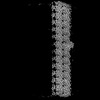
| Projections & slices | Image control
Images are generated by Spider. | ||||||||||||||||||||||||||||||||||||
| Voxel size | X=Y=Z: 1.245 Å | ||||||||||||||||||||||||||||||||||||
| Density |
| ||||||||||||||||||||||||||||||||||||
| Symmetry | Space group: 1 | ||||||||||||||||||||||||||||||||||||
| Details | EMDB XML:
|
 Z (Sec.)
 Y (Row.)
 X (Col.)


-Supplemental data
-Mask #1
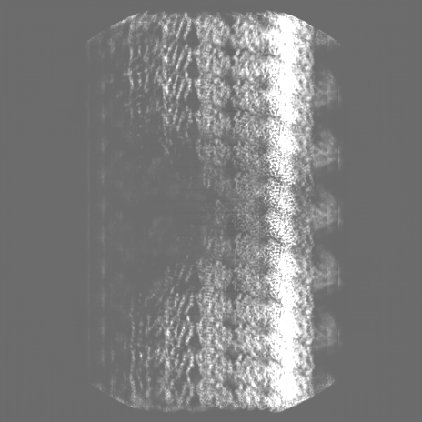
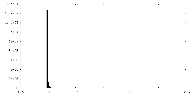
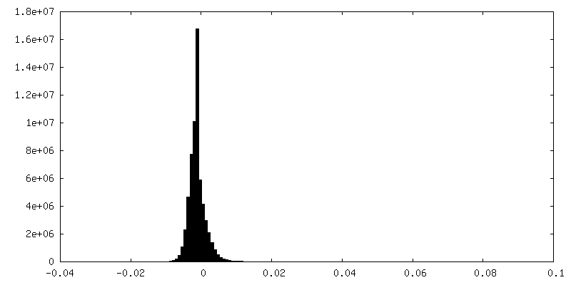
| File | emd_42948_msk_1.map | ||||||||||||
|---|---|---|---|---|---|---|---|---|---|---|---|---|---|
| Projections & Slices |
| ||||||||||||
| Density Histograms |


-Additional map: Protofilament reconstruction in class 1, raw full map
| File | emd_42948_additional_1.map | ||||||||||||
|---|---|---|---|---|---|---|---|---|---|---|---|---|---|
| Annotation | Protofilament reconstruction in class 1, raw full map | ||||||||||||
| Projections & Slices |
| ||||||||||||
| Density Histograms |


-Additional map: Protofilament reconstruction in class 1, DeepEMhancer processed
| File | emd_42948_additional_2.map | ||||||||||||
|---|---|---|---|---|---|---|---|---|---|---|---|---|---|
| Annotation | Protofilament reconstruction in class 1, DeepEMhancer processed | ||||||||||||
| Projections & Slices |
| ||||||||||||
| Density Histograms |


-Half map: Protofilament reconstruction in class 1, raw half map
| File | emd_42948_half_map_1.map | ||||||||||||
|---|---|---|---|---|---|---|---|---|---|---|---|---|---|
| Annotation | Protofilament reconstruction in class 1, raw half map | ||||||||||||
| Projections & Slices |
| ||||||||||||
| Density Histograms |


-Half map: Protofilament reconstruction in class 1, raw half map
| File | emd_42948_half_map_2.map | ||||||||||||
|---|---|---|---|---|---|---|---|---|---|---|---|---|---|
| Annotation | Protofilament reconstruction in class 1, raw half map | ||||||||||||
| Projections & Slices |
| ||||||||||||
| Density Histograms |


- Sample components
-Entire : Microtubule protofilament
| Entire | Name: Microtubule protofilament |
|---|---|
| Components |
|
-Supramolecule #1: Microtubule protofilament
| Supramolecule | Name: Microtubule protofilament / type: complex / ID: 1 / Parent: 0 Details: Protofilament reconstruction of CCP5 decorated microtubules |
|---|
-Supramolecule #2: Microtubule protofilament
| Supramolecule | Name: Microtubule protofilament / type: complex / ID: 2 / Parent: 1 |
|---|---|
| Source (natural) | Organism: |
-Experimental details
-Structure determination
| Method | cryo EM |
|---|---|
 Processing Processing | single particle reconstruction |
| Aggregation state | filament |
-Sample preparation
| Concentration | 0.1 mg/mL | ||||||||||||
|---|---|---|---|---|---|---|---|---|---|---|---|---|---|
| Buffer | pH: 7.4 Component:
| ||||||||||||
| Grid | Model: Au-flat 1.2/1.3 / Material: GOLD / Mesh: 300 / Support film - Material: GOLD / Support film - topology: HOLEY / Support film - Film thickness: 45 / Pretreatment - Type: GLOW DISCHARGE / Pretreatment - Time: 30 sec. | ||||||||||||
| Vitrification | Cryogen name: ETHANE / Chamber humidity: 90 % / Chamber temperature: 303 K / Instrument: LEICA EM GP | ||||||||||||
| Details | Pig brain microtubules double cycled with GMPCPP and decorated with human CCP5. |
- Electron microscopy
| Microscope | FEI TITAN KRIOS |
|---|---|
| Specialist optics | Energy filter - Slit width: 20 eV |
| Image recording | Film or detector model: GATAN K3 (6k x 4k) / Average exposure time: 1.651 sec. / Average electron dose: 53.34 e/Å2 |
| Electron beam | Acceleration voltage: 300 kV / Electron source:  FIELD EMISSION GUN FIELD EMISSION GUN |
| Electron optics | C2 aperture diameter: 70.0 µm / Illumination mode: FLOOD BEAM / Imaging mode: BRIGHT FIELD / Cs: 2.7 mm / Nominal defocus max: 2.2 µm / Nominal defocus min: 0.8 µm / Nominal magnification: 135000 |
| Sample stage | Cooling holder cryogen: NITROGEN |
| Experimental equipment |  Model: Titan Krios / Image courtesy: FEI Company |
+Image processing

-Atomic model buiding 1
| Initial model | PDB ID: Chain - Source name: PDB / Chain - Initial model type: experimental model |
|---|