 Movie
Movie Controller

[English] 日本語
 Yorodumi
- PDB-8us2: P22121 Crystal structure of TamA from Pseudomonas aeruginosa at 3... -
+ Open data

- Basic information
| Entry | Database: PDB / ID: 8us2 | ||||||
|---|---|---|---|---|---|---|---|
| Title | P22121 Crystal structure of TamA from Pseudomonas aeruginosa at 3.95 Ang | ||||||
 Components Components | Translocation and assembly module subunit TamA | ||||||
 Keywords Keywords | MEMBRANE PROTEIN / Omp85 / protein translocation | ||||||
| Function / homology |  Function and homology information Function and homology information | ||||||
| Biological species |   Pseudomonas aeruginosa (bacteria) Pseudomonas aeruginosa (bacteria) | ||||||
| Method |  X-RAY DIFFRACTION / SYNCHROTRON / MOLECULAR REPLACEMENT / Resolution: 3.955 Å X-RAY DIFFRACTION / SYNCHROTRON / MOLECULAR REPLACEMENT / Resolution: 3.955 Å | ||||||
 Authors Authors | Mellouk, A. / Moraes, T.F. / Calmettes, C. | ||||||
| Funding support |  Canada, 1items Canada, 1items
| ||||||
 Citation Citation | Journal: Proc.Natl.Acad.Sci.USA / Year: 2024 Title: POTRA domains of the TamA insertase interact with the outer membrane and modulate membrane properties. Authors: Mellouk, A. / Jaouen, P. / Ruel, L.J. / Le, M. / Martini, C. / Moraes, T.F. / El Bakkouri, M. / Lague, P. / Boisselier, E. / Calmettes, C. | ||||||
| History |
|
- Structure visualization
| Structure viewer | Molecule: MolmilJmol/JSmol |
|---|
- Downloads & links
-Download
| PDBx/mmCIF format | 8us2.cif.gz | 210 KB | Display | PDBx/mmCIF format |
|---|---|---|---|---|
| PDB format | pdb8us2.ent.gz | 168.8 KB | Display | PDB format |
| PDBx/mmJSON format | 8us2.json.gz | Tree view | PDBx/mmJSON format | |
| Others |  Other downloads Other downloads |
-Validation report
| Summary document | 8us2_validation.pdf.gz | 440.5 KB | Display | wwPDB validaton report |
|---|---|---|---|---|
| Full document | 8us2_full_validation.pdf.gz | 463.1 KB | Display | |
| Data in XML | 8us2_validation.xml.gz | 38.3 KB | Display | |
| Data in CIF | 8us2_validation.cif.gz | 51 KB | Display | |
| Arichive directory | https://data.pdbj.org/pub/pdb/validation_reports/us/8us2ftp://data.pdbj.org/pub/pdb/validation_reports/us/8us2 | HTTPS FTP |
-Related structure data
-Links
 PDBj
PDBj- Assembly
| Deposited unit | 
| ||||||||
|---|---|---|---|---|---|---|---|---|---|
| 1 | 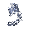
| ||||||||
| 2 | 
| ||||||||
| Unit cell |
|
-Components
| #1: Protein | Mass: 61651.949 Da / Num. of mol.: 2 / Fragment: Mature TamA Source method: isolated from a genetically manipulated source Source: (gene. exp.) Pseudomonas aeruginosa (bacteria)Strain: ATCC 15692 / DSM 22644 / CIP 104116 / JCM 14847 / LMG 12228 / 1C / PRS 101 / PAO1 Gene: PA2543 / Plasmid: pET / Production host: |
|---|
-Experimental details
-Experiment
| Experiment | Method: X-RAY DIFFRACTION / Number of used crystals: 1 |
|---|
- Sample preparation
| Crystal | Density Matthews: 3.62 Å3/Da / Density % sol: 65.99 % |
|---|---|
| Crystal grow | Temperature: 293 K / Method: vapor diffusion, hanging drop / pH: 7.2 Details: 12% peg 4K, 50 mM MgCl2, 70 mM Hepes pH 7.2, 4% isopropanol, 10% glycerol |
-Data collection
| Diffraction | Mean temperature: 77 K / Serial crystal experiment: N | ||||||||||||||||||||||||||||||||||||||||||||||||||||||||||||||||||||||||||||||||||||||||
|---|---|---|---|---|---|---|---|---|---|---|---|---|---|---|---|---|---|---|---|---|---|---|---|---|---|---|---|---|---|---|---|---|---|---|---|---|---|---|---|---|---|---|---|---|---|---|---|---|---|---|---|---|---|---|---|---|---|---|---|---|---|---|---|---|---|---|---|---|---|---|---|---|---|---|---|---|---|---|---|---|---|---|---|---|---|---|---|---|---|
| Diffraction source | Source: SYNCHROTRON / Site: APS  / Beamline: 24-ID-C / Wavelength: 0.97910 Ang / Beamline: 24-ID-C / Wavelength: 0.97910 Ang | ||||||||||||||||||||||||||||||||||||||||||||||||||||||||||||||||||||||||||||||||||||||||
| Detector | Type: ADSC QUANTUM 315 / Detector: CCD / Date: Oct 12, 2013 | ||||||||||||||||||||||||||||||||||||||||||||||||||||||||||||||||||||||||||||||||||||||||
| Radiation | Protocol: SINGLE WAVELENGTH / Monochromatic (M) / Laue (L): M / Scattering type: x-ray | ||||||||||||||||||||||||||||||||||||||||||||||||||||||||||||||||||||||||||||||||||||||||
| Radiation wavelength | Wavelength: 0.9791 Å / Relative weight: 1 | ||||||||||||||||||||||||||||||||||||||||||||||||||||||||||||||||||||||||||||||||||||||||
| Reflection | Resolution: 3.955→44.063 Å / Num. all: 16231 / Num. obs: 16231 / % possible obs: 99.7 % / Redundancy: 3.9 % / Biso Wilson estimate: 123.22 Å2 / Rpim(I) all: 0.155 / Rrim(I) all: 0.309 / Rsym value: 0.222 / Net I/av σ(I): 2.739 / Net I/σ(I): 7.2 / Num. measured all: 62873 | ||||||||||||||||||||||||||||||||||||||||||||||||||||||||||||||||||||||||||||||||||||||||
| Reflection shell | Diffraction-ID: 1
|
- Processing
| Software |
| |||||||||||||||||||||||||||||||||||||||||||||||||
|---|---|---|---|---|---|---|---|---|---|---|---|---|---|---|---|---|---|---|---|---|---|---|---|---|---|---|---|---|---|---|---|---|---|---|---|---|---|---|---|---|---|---|---|---|---|---|---|---|---|---|
| Refinement | Method to determine structure: MOLECULAR REPLACEMENT / Resolution: 3.955→39.255 Å / SU ML: 0.87 / Cross valid method: FREE R-VALUE / σ(F): 1.34 / Phase error: 45.57 / Stereochemistry target values: ML
| |||||||||||||||||||||||||||||||||||||||||||||||||
| Solvent computation | Shrinkage radii: 0.9 Å / VDW probe radii: 1.11 Å / Solvent model: FLAT BULK SOLVENT MODEL | |||||||||||||||||||||||||||||||||||||||||||||||||
| Displacement parameters | Biso max: 550 Å2 / Biso mean: 159.9052 Å2 / Biso min: 6.18 Å2 | |||||||||||||||||||||||||||||||||||||||||||||||||
| Refinement step | Cycle: final / Resolution: 3.955→39.255 Å
| |||||||||||||||||||||||||||||||||||||||||||||||||
| Refine LS restraints |
| |||||||||||||||||||||||||||||||||||||||||||||||||
| LS refinement shell |
|
