 Movie
Movie Controller

[English] 日本語
 Yorodumi
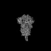
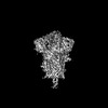
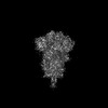
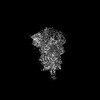
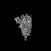
- PDB-8uir: SARS-CoV-2 Omicron-XBB.1.16 3-RBD down Spike Protein Trimer conse... -
+ Open data

- Basic information
| Entry | Database: PDB / ID: 8uir | ||||||
|---|---|---|---|---|---|---|---|
| Title | SARS-CoV-2 Omicron-XBB.1.16 3-RBD down Spike Protein Trimer consensus (S-GSAS-Omicron-XBB.1.16) | ||||||
 Components Components | Spike glycoprotein | ||||||
 Keywords Keywords | VIRAL PROTEIN / SARS-COV-2 / Glycoprotein / Trimer | ||||||
| Function / homology |  Function and homology information Function and homology informationsymbiont-mediated disruption of host tissue / Maturation of spike protein / Translation of Structural Proteins / Virion Assembly and Release / host cell surface / host extracellular region / symbiont-mediated-mediated suppression of host tetherin activity / Induction of Cell-Cell Fusion / structural constituent of virion / positive regulation of viral entry into host cell ...symbiont-mediated disruption of host tissue / Maturation of spike protein / Translation of Structural Proteins / Virion Assembly and Release / host cell surface / host extracellular region / symbiont-mediated-mediated suppression of host tetherin activity / Induction of Cell-Cell Fusion / structural constituent of virion / positive regulation of viral entry into host cell / membrane fusion / host cell endoplasmic reticulum-Golgi intermediate compartment membrane / Attachment and Entry / entry receptor-mediated virion attachment to host cell / receptor-mediated virion attachment to host cell / host cell surface receptor binding / symbiont-mediated suppression of host innate immune response / endocytosis involved in viral entry into host cell / receptor ligand activity / fusion of virus membrane with host plasma membrane / fusion of virus membrane with host endosome membrane / viral envelope / symbiont entry into host cell / virion attachment to host cell / host cell plasma membrane / SARS-CoV-2 activates/modulates innate and adaptive immune responses / virion membrane / membrane / identical protein binding / plasma membrane Similarity search - Function | ||||||
| Biological species |   Severe acute respiratory syndrome coronavirus 2 Severe acute respiratory syndrome coronavirus 2 | ||||||
| Method | ELECTRON MICROSCOPY / single particle reconstruction / cryo EM / Resolution: 3.2 Å | ||||||
 Authors Authors | Zhang, Q.E. / Acharya, P. | ||||||
| Funding support |  United States, 1items United States, 1items
| ||||||
 Citation Citation | Journal: Mol Cell / Year: 2024 Title: SARS-CoV-2 Omicron XBB lineage spike structures, conformations, antigenicity, and receptor recognition. Authors: Qianyi E Zhang / Jared Lindenberger / Ruth J Parsons / Bhishem Thakur / Rob Parks / Chan Soo Park / Xiao Huang / Salam Sammour / Katarzyna Janowska / Taylor N Spence / Robert J Edwards / ...Authors: Qianyi E Zhang / Jared Lindenberger / Ruth J Parsons / Bhishem Thakur / Rob Parks / Chan Soo Park / Xiao Huang / Salam Sammour / Katarzyna Janowska / Taylor N Spence / Robert J Edwards / Mitchell Martin / Wilton B Williams / Sophie Gobeil / David C Montefiori / Bette Korber / Kevin O Saunders / Barton F Haynes / Rory Henderson / Priyamvada Acharya /  Abstract: A recombinant lineage of the severe acute respiratory syndrome coronavirus 2 (SARS-CoV-2) Omicron variant, named XBB, appeared in late 2022 and evolved descendants that successively swept local and ...A recombinant lineage of the severe acute respiratory syndrome coronavirus 2 (SARS-CoV-2) Omicron variant, named XBB, appeared in late 2022 and evolved descendants that successively swept local and global populations. XBB lineage members were noted for their improved immune evasion and transmissibility. Here, we determine cryoelectron microscopy (cryo-EM) structures of XBB.1.5, XBB.1.16, EG.5, and EG.5.1 spike (S) ectodomains to reveal reinforced 3-receptor binding domain (RBD)-down receptor-inaccessible closed states mediated by interprotomer RBD interactions previously observed in BA.1 and BA.2. Improved XBB.1.5 and XBB.1.16 RBD stability compensated for stability loss caused by early Omicron mutations, while the F456L substitution reduced EG.5 RBD stability. S1 subunit mutations had long-range impacts on conformation and epitope presentation in the S2 subunit. Our results reveal continued S protein evolution via simultaneous optimization of multiple parameters, including stability, receptor binding, and immune evasion, and the dramatic effects of relatively few residue substitutions in altering the S protein conformational landscape. | ||||||
| History |
|
- Structure visualization
| Structure viewer | Molecule: MolmilJmol/JSmol |
|---|
- Downloads & links
-Download
| PDBx/mmCIF format | 8uir.cif.gz | 634.6 KB | Display | PDBx/mmCIF format |
|---|---|---|---|---|
| PDB format | pdb8uir.ent.gz | 509 KB | Display | PDB format |
| PDBx/mmJSON format | 8uir.json.gz | Tree view | PDBx/mmJSON format | |
| Others |  Other downloads Other downloads |
-Validation report
| Arichive directory | https://data.pdbj.org/pub/pdb/validation_reports/ui/8uirftp://data.pdbj.org/pub/pdb/validation_reports/ui/8uir | HTTPS FTP |
|---|
-Related structure data
| Related structure data | 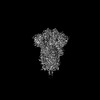 42302MC  8uk1C  8ukdC  8ukfC  8v0lC 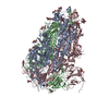 8v0mC  8v0nC  8v0oC 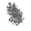 8v0pC  8v0qC  8v0rC  8v0sC 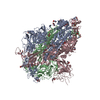 8v0tC  8v0uC  8v0vC  8v0wC  8v0xC  9aywC  9ayxC  9ayyC M: map data used to model this data C: citing same article ( |
|---|---|
| Similar structure data |


-Links
 PDBj
PDBj


- Assembly
| Deposited unit | 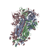
|
|---|---|
| 1 |
|
-Components
| #1: Protein | Mass: 142230.578 Da / Num. of mol.: 3 / Mutation: residues 682-685 mutated from RRAR to GSAS Source method: isolated from a genetically manipulated source Source: (gene. exp.) Severe acute respiratory syndrome coronavirus 2Gene: S, 2 / Production host:  Homo sapiens (human) / References: UniProt: P0DTC2 Homo sapiens (human) / References: UniProt: P0DTC2#2: Polysaccharide | 2-acetamido-2-deoxy-beta-D-glucopyranose-(1-4)-2-acetamido-2-deoxy-beta-D-glucopyranose Source method: isolated from a genetically manipulated source #3: Sugar | ChemComp-NAG /   Type: D-saccharide, beta linking / Mass: 221.208 Da / Num. of mol.: 30 Type: D-saccharide, beta linking / Mass: 221.208 Da / Num. of mol.: 30Source method: isolated from a genetically manipulated source Formula: C8H15NO6 / Feature type: SUBJECT OF INVESTIGATION Has ligand of interest | Y | Has protein modification | Y | |
|---|
-Experimental details
-Experiment
| Experiment | Method: ELECTRON MICROSCOPY |
|---|---|
| EM experiment | Aggregation state: PARTICLE / 3D reconstruction method: single particle reconstruction |
- Sample preparation
| Component | Name: Cryo-EM structure of GSAS-XBB.1.16 / Type: COMPLEX / Entity ID: #1 / Source: RECOMBINANT |
|---|---|
| Source (natural) | Organism: Severe acute respiratory syndrome coronavirus 2 |
| Source (recombinant) | Organism: Homo sapiens (human) |
| Buffer solution | pH: 8 |
| Specimen | Conc.: 2 mg/ml / Embedding applied: NO / Shadowing applied: NO / Staining applied: NO / Vitrification applied: YES |
| Specimen support | Grid material: COPPER / Grid mesh size: 300 divisions/in. / Grid type: Quantifoil R1.2/1.3 |
| Vitrification | Instrument: LEICA EM GP / Cryogen name: ETHANE / Humidity: 95 % / Chamber temperature: 295.15 K |
- Electron microscopy imaging
| Experimental equipment |  Model: Titan Krios / Image courtesy: FEI Company |
|---|---|
| Microscopy | Model: FEI TITAN KRIOS |
| Electron gun | Electron source:  FIELD EMISSION GUN / Accelerating voltage: 300 kV / Illumination mode: FLOOD BEAM FIELD EMISSION GUN / Accelerating voltage: 300 kV / Illumination mode: FLOOD BEAM |
| Electron lens | Mode: BRIGHT FIELD / Nominal magnification: 81000 X / Nominal defocus max: 5000 nm / Nominal defocus min: 350 nm / Cs: 2.7 mm |
| Specimen holder | Cryogen: NITROGEN |
| Image recording | Electron dose: 59 e/Å2 / Film or detector model: GATAN K3 BIOQUANTUM (6k x 4k) / Num. of real images: 6629 |
| Image scans | Width: 5760 / Height: 4092 |
- Processing
| EM software |
| ||||||||||||||||
|---|---|---|---|---|---|---|---|---|---|---|---|---|---|---|---|---|---|
| CTF correction | Type: PHASE FLIPPING AND AMPLITUDE CORRECTION | ||||||||||||||||
| Particle selection | Num. of particles selected: 1284770 | ||||||||||||||||
| 3D reconstruction | Resolution: 3.2 Å / Resolution method: FSC 0.143 CUT-OFF / Num. of particles: 852149 / Symmetry type: POINT | ||||||||||||||||
| Atomic model building | Protocol: FLEXIBLE FIT / Space: REAL |
