 Movie
Movie Controller

[English] 日本語
 Yorodumi
- EMDB-43463: SARS-CoV-2 Omicron-EG.5 1-RBD up Spike Protein Trimer (S-GSAS-Omi... -
+ Open data

- Basic information
| Entry |  | |||||||||
|---|---|---|---|---|---|---|---|---|---|---|
| Title | SARS-CoV-2 Omicron-EG.5 1-RBD up Spike Protein Trimer (S-GSAS-Omicron-EG.5) | |||||||||
 Map data Map data | ||||||||||
 Sample Sample |
| |||||||||
 Keywords Keywords | SARS-COV-2 / Glycoprotein / Trimer / VIRAL PROTEIN | |||||||||
| Biological species |   Severe acute respiratory syndrome coronavirus 2 Severe acute respiratory syndrome coronavirus 2 | |||||||||
| Method | single particle reconstruction / cryo EM / Resolution: 3.4 Å | |||||||||
 Authors Authors | Zhang QE / Acharya P | |||||||||
| Funding support |  United States, 1 items United States, 1 items
| |||||||||
 Citation Citation | Journal: Mol Cell / Year: 2024 Title: SARS-CoV-2 Omicron XBB lineage spike structures, conformations, antigenicity, and receptor recognition. Authors: Qianyi E Zhang / Jared Lindenberger / Ruth J Parsons / Bhishem Thakur / Rob Parks / Chan Soo Park / Xiao Huang / Salam Sammour / Katarzyna Janowska / Taylor N Spence / Robert J Edwards / ...Authors: Qianyi E Zhang / Jared Lindenberger / Ruth J Parsons / Bhishem Thakur / Rob Parks / Chan Soo Park / Xiao Huang / Salam Sammour / Katarzyna Janowska / Taylor N Spence / Robert J Edwards / Mitchell Martin / Wilton B Williams / Sophie Gobeil / David C Montefiori / Bette Korber / Kevin O Saunders / Barton F Haynes / Rory Henderson / Priyamvada Acharya /  Abstract: A recombinant lineage of the severe acute respiratory syndrome coronavirus 2 (SARS-CoV-2) Omicron variant, named XBB, appeared in late 2022 and evolved descendants that successively swept local and ...A recombinant lineage of the severe acute respiratory syndrome coronavirus 2 (SARS-CoV-2) Omicron variant, named XBB, appeared in late 2022 and evolved descendants that successively swept local and global populations. XBB lineage members were noted for their improved immune evasion and transmissibility. Here, we determine cryoelectron microscopy (cryo-EM) structures of XBB.1.5, XBB.1.16, EG.5, and EG.5.1 spike (S) ectodomains to reveal reinforced 3-receptor binding domain (RBD)-down receptor-inaccessible closed states mediated by interprotomer RBD interactions previously observed in BA.1 and BA.2. Improved XBB.1.5 and XBB.1.16 RBD stability compensated for stability loss caused by early Omicron mutations, while the F456L substitution reduced EG.5 RBD stability. S1 subunit mutations had long-range impacts on conformation and epitope presentation in the S2 subunit. Our results reveal continued S protein evolution via simultaneous optimization of multiple parameters, including stability, receptor binding, and immune evasion, and the dramatic effects of relatively few residue substitutions in altering the S protein conformational landscape. | |||||||||
| History |
|
- Structure visualization
| Supplemental images |
|---|

- Downloads & links
-EMDB archive
| Map data | emd_43463.map.gz | 115.9 MB |  EMDB map data format EMDB map data format | |
|---|---|---|---|---|
| Header (meta data) | emd-43463-v30.xmlemd-43463.xml | 16.5 KB 16.5 KB | Display Display | EMDB header |
| FSC (resolution estimation) | emd_43463_fsc.xml | 11.9 KB | Display | FSC data file |
| Images |  emd_43463.png emd_43463.png | 126.6 KB | ||
| Filedesc metadata | emd-43463.cif.gz | 5.9 KB | ||
| Others | emd_43463_half_map_1.map.gzemd_43463_half_map_2.map.gz | 114 MB 114 MB | ||
| Archive directory |  http://ftp.pdbj.org/pub/emdb/structures/EMD-43463ftp://ftp.pdbj.org/pub/emdb/structures/EMD-43463 http://ftp.pdbj.org/pub/emdb/structures/EMD-43463ftp://ftp.pdbj.org/pub/emdb/structures/EMD-43463 | HTTPS FTP |
-Related structure data
| Related structure data | 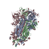 8uirC  8uk1C 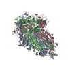 8ukdC 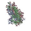 8ukfC 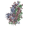 8v0lC  8v0mC  8v0nC  8v0oC  8v0pC  8v0qC  8v0rC  8v0sC 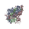 8v0tC  8v0uC  8v0vC  8v0wC  8v0xC  9aywC  9ayxC  9ayyC C: citing same article ( |
|---|


-Links
| EMDB pages | EMDB (EBI/PDBe) / EMDataResource |
|---|
-Map
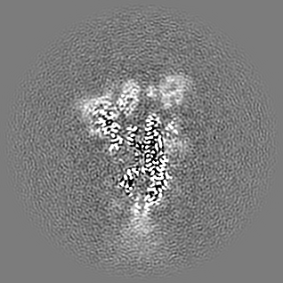
| File | Download / File: emd_43463.map.gz / Format: CCP4 / Size: 122.7 MB / Type: IMAGE STORED AS FLOATING POINT NUMBER (4 BYTES) | ||||||||||||||||||||||||||||||||||||
|---|---|---|---|---|---|---|---|---|---|---|---|---|---|---|---|---|---|---|---|---|---|---|---|---|---|---|---|---|---|---|---|---|---|---|---|---|---|
| Projections & slices | Image control
Images are generated by Spider. | ||||||||||||||||||||||||||||||||||||
| Voxel size | X=Y=Z: 1.08 Å | ||||||||||||||||||||||||||||||||||||
| Density |
| ||||||||||||||||||||||||||||||||||||
| Symmetry | Space group: 1 | ||||||||||||||||||||||||||||||||||||
| Details | EMDB XML:
|
 Z (Sec.)
 Y (Row.)
 X (Col.)


-Supplemental data
-Half map: #1
| File | emd_43463_half_map_1.map | ||||||||||||
|---|---|---|---|---|---|---|---|---|---|---|---|---|---|
| Projections & Slices |
| ||||||||||||
| Density Histograms |


-Half map: #2
| File | emd_43463_half_map_2.map | ||||||||||||
|---|---|---|---|---|---|---|---|---|---|---|---|---|---|
| Projections & Slices |
| ||||||||||||
| Density Histograms |


- Sample components
-Entire : GSAS-EG.5
| Entire | Name: GSAS-EG.5 |
|---|---|
| Components |
|
-Supramolecule #1: GSAS-EG.5
| Supramolecule | Name: GSAS-EG.5 / type: complex / ID: 1 / Parent: 0 / Macromolecule list: all |
|---|---|
| Source (natural) | Organism: Severe acute respiratory syndrome coronavirus 2 |
-Macromolecule #1: Spike glycoprotein
| Macromolecule | Name: Spike glycoprotein / type: protein_or_peptide / ID: 1 / Enantiomer: LEVO |
|---|---|
| Source (natural) | Organism: Severe acute respiratory syndrome coronavirus 2 |
| Recombinant expression | Organism:  Homo sapiens (human) Homo sapiens (human) |
| Sequence | String: MFVFLVLLPL VSSQCVNLIT RTQSYTNSFT RGVYYPDKVF RSSVLHSTQD LFLPFFSNVT WFHAIHVSGT NGTKRFDNPA LPFNDGVYF ASTEKSNIIR GWIFGTTLDS KTQSLLIVNN ATNVVIKVCE FQFCNDPFLD VYQKNNKSWM ESEFRVYSSA N NCTFEYVS ...String: MFVFLVLLPL VSSQCVNLIT RTQSYTNSFT RGVYYPDKVF RSSVLHSTQD LFLPFFSNVT WFHAIHVSGT NGTKRFDNPA LPFNDGVYF ASTEKSNIIR GWIFGTTLDS KTQSLLIVNN ATNVVIKVCE FQFCNDPFLD VYQKNNKSWM ESEFRVYSSA N NCTFEYVS QPFLMDLEGK EGNFKNLREF VFKNIDGYFK IYSKHTPINL ERDLPQGFSA LEPLVDLPIG INITRFQTLL AL HRSYLTP VDSSSGWTAG AAAYYVGYLQ PRTFLLKYNE NGTITDAVDC ALDPLSETKC TLKSFTVEKG IYQTSNFRVQ PTE SIVRFP NITNLCPFHE VFNATTFASV YAWNRKRISN CVADYSVIYN FAPFFAFKCY GVSPTKLNDL CFTNVYADSF VIRG NEVSQ IAPGQTGNIA DYNYKLPDDF TGCVIAWNSN KLDSKPSGNY NYLYRLLRKS KLKPFERDIS TEIYQAGNKP CNGVA GPNC YSPLQSYGFR PTYGVGHQPY RVVVLSFELL HAPATVCGPK KSTNLVKNKC VNFNFNGLTG TGVLTESNKK FLPFQQ FGR DIADTTDAVR DPQTLEILDI TPCSFGGVSV ITPGTNTSNQ VAVLYQGVNC TEVPVAIHAD QLTPTWRVYS TGSNVFQ TR AGCLIGAEYV NNSYECDIPI GAGICASYQT QTKSHGSASS VASQSIIAYT MSLGAENSVA YSNNSIAIPT NFTISVTT E ILPVSMTKTS VDCTMYICGD STECSNLLLQ YGSFCTQLKR ALTGIAVEQD KNTQEVFAQV KQIYKTPPIK YFGGFNFSQ ILPDPSKPSK RSFIEDLLFN KVTLADAGFI KQYGDCLGDI AARDLICAQK FNGLTVLPPL LTDEMIAQYT SALLAGTITS GWTFGAGAA LQIPFAMQMA YRFNGIGVTQ NVLYENQKLI ANQFNSAIGK IQDSLSSTAS ALGKLQDVVN HNAQALNTLV K QLSSKFGA ISSVLNDILS RLDKVEAEVQ IDRLITGRLQ SLQTYVTQQL IRAAEIRASA NLAATKMSEC VLGQSKRVDF CG KGYHLMS FPQSAPHGVV FLHVTYVPAQ EKNFTTAPAI CHDGKAHFPR EGVFVSNGTH WFVTQRNFYE PQIITTDNTF VSG NCDVVI GIVNNTVYDP LQPELDSFKE ELDKYFKNHT SPDVDLGDIS GINASVVNIQ KEIDRLNEVA KNLNESLIDL QELG KYEQG SGYIPEAPRD GQAYVRKDGE WVLLSTFLGR SLEVLFQGPG HHHHHHHHSA WSHPQFEKGG GSGGGGSGGS AWSHP QFEK |
-Experimental details
-Structure determination
| Method | cryo EM |
|---|---|
 Processing Processing | single particle reconstruction |
| Aggregation state | particle |
-Sample preparation
| Concentration | 1.5 mg/mL |
|---|---|
| Buffer | pH: 8 |
| Grid | Model: Quantifoil R1.2/1.3 / Material: COPPER / Mesh: 300 / Support film - Material: CARBON / Support film - topology: HOLEY / Pretreatment - Type: GLOW DISCHARGE / Pretreatment - Time: 15 sec. |
| Vitrification | Cryogen name: ETHANE / Chamber humidity: 95 % / Chamber temperature: 295.15 K / Instrument: LEICA EM GP |
- Electron microscopy
| Microscope | FEI TITAN KRIOS |
|---|---|
| Image recording | Film or detector model: GATAN K3 BIOQUANTUM (6k x 4k) / Digitization - Dimensions - Width: 5760 pixel / Digitization - Dimensions - Height: 4092 pixel / Number real images: 8079 / Average electron dose: 72.9 e/Å2 |
| Electron beam | Acceleration voltage: 300 kV / Electron source:  FIELD EMISSION GUN FIELD EMISSION GUN |
| Electron optics | Illumination mode: FLOOD BEAM / Imaging mode: BRIGHT FIELD / Cs: 2.7 mm / Nominal defocus max: 5.0 µm / Nominal defocus min: 0.35000000000000003 µm / Nominal magnification: 81000 |
| Sample stage | Cooling holder cryogen: NITROGEN |
| Experimental equipment |  Model: Titan Krios / Image courtesy: FEI Company |
+Image processing

-Atomic model buiding 1
| Refinement | Space: REAL / Protocol: FLEXIBLE FIT |
|---|
