 Movie
Movie Controller


+ Open data

- Basic information
| Entry | Database: PDB / ID: 8tzh | ||||||
|---|---|---|---|---|---|---|---|
| Title | Structure of full-length LRRK2 bound to MLi-2 (I2020T mutant) | ||||||
 Components Components |
| ||||||
 Keywords Keywords | PROTEIN BINDING / GtPase / kinase / inhibitors | ||||||
| Function / homology |  Function and homology information Function and homology informationcaveola neck / : / beta-catenin destruction complex binding / regulation of branching morphogenesis of a nerve / Wnt signalosome assembly / negative regulation of motile cilium assembly / regulation of kidney size / regulation of cell projection organization / tangential migration from the subventricular zone to the olfactory bulb / GTP-dependent protein kinase activity ...caveola neck / : / beta-catenin destruction complex binding / regulation of branching morphogenesis of a nerve / Wnt signalosome assembly / negative regulation of motile cilium assembly / regulation of kidney size / regulation of cell projection organization / tangential migration from the subventricular zone to the olfactory bulb / GTP-dependent protein kinase activity / regulation of SNARE complex assembly / regulation of neuroblast proliferation / regulation of ER to Golgi vesicle-mediated transport / protein localization to endoplasmic reticulum exit site / negative regulation of late endosome to lysosome transport / regulation of mitochondrial depolarization / : / peroxidase inhibitor activity / positive regulation of dopamine receptor signaling pathway / amphisome / regulation of synaptic vesicle transport / : / regulation of CAMKK-AMPK signaling cascade / co-receptor binding / negative regulation of GTPase activity / regulation of dopamine receptor signaling pathway / positive regulation of microglial cell activation / regulation of retrograde transport, endosome to Golgi / regulation of neuron maturation / cellular response to curcumin / olfactory bulb development / cytoplasmic side of mitochondrial outer membrane / positive regulation of synaptic vesicle endocytosis / JUN kinase kinase kinase activity / negative regulation of autophagosome assembly / multivesicular body, internal vesicle / negative regulation of excitatory postsynaptic potential / regulation of cAMP/PKA signal transduction / striatum development / neuron projection arborization / regulation of dendritic spine morphogenesis / mitochondrion localization / protein localization to mitochondrion / cellular response to dopamine / positive regulation of mitochondrial outer membrane permeabilization involved in apoptotic signaling pathway / endoplasmic reticulum organization / negative regulation of protein processing / positive regulation of protein autoubiquitination / Wnt signalosome / positive regulation of programmed cell death / GTP metabolic process / regulation of canonical Wnt signaling pathway / syntaxin-1 binding / regulation of reactive oxygen species metabolic process / Golgi-associated vesicle / PTK6 promotes HIF1A stabilization / lysosome organization / clathrin binding / negative regulation of macroautophagy / regulation of mitochondrial fission / Lewy body / regulation of synaptic vesicle exocytosis / regulation of locomotion / intracellular distribution of mitochondria / neuromuscular junction development / Golgi organization / protein kinase A binding / microvillus / exploration behavior / autolysosome / locomotory exploration behavior / endoplasmic reticulum exit site / negative regulation of Notch signaling pathway / MAP kinase kinase kinase activity / canonical Wnt signaling pathway / regulation of synaptic vesicle endocytosis / negative regulation of endoplasmic reticulum stress-induced intrinsic apoptotic signaling pathway / Rho protein signal transduction / regulation of synaptic transmission, glutamatergic / presynaptic cytosol / JNK cascade / cellular response to manganese ion / phagocytic vesicle / neuron projection morphogenesis / positive regulation of autophagy / dendrite cytoplasm / cellular response to starvation / positive regulation of protein ubiquitination / determination of adult lifespan / GTPase activator activity / regulation of autophagy / SNARE binding / cellular response to reactive oxygen species / mitochondrion organization / trans-Golgi network / calcium-mediated signaling / regulation of membrane potential / regulation of protein stability / excitatory postsynaptic potential / tubulin binding Similarity search - Function | ||||||
| Biological species | synthetic construct (others) Homo sapiens (human) Homo sapiens (human) | ||||||
| Method | ELECTRON MICROSCOPY / single particle reconstruction / cryo EM / Resolution: 3.9 Å | ||||||
 Authors Authors | Sanz-Murillo, M. / Villagran-Suarez, A. / Alegrio Louro, J. / Leschziner, A. | ||||||
| Funding support |  United States, 1items United States, 1items
| ||||||
 Citation Citation | Journal: Sci Adv / Year: 2023 Title: Inhibition of Parkinson's disease-related LRRK2 by type I and type II kinase inhibitors: Activity and structures. Authors: Marta Sanz Murillo / Amalia Villagran Suarez / Verena Dederer / Deep Chatterjee / Jaime Alegrio Louro / Stefan Knapp / Sebastian Mathea / Andres E Leschziner /  Abstract: Mutations in leucine-rich repeat kinase 2 (LRRK2) are a common cause of familial Parkinson's disease (PD) and a risk factor for the sporadic form. Increased kinase activity was shown in patients with ...Mutations in leucine-rich repeat kinase 2 (LRRK2) are a common cause of familial Parkinson's disease (PD) and a risk factor for the sporadic form. Increased kinase activity was shown in patients with both familial and sporadic PD, making LRRK2 kinase inhibitors a major focus of drug development efforts. Although much progress has been made in understanding the structural biology of LRRK2, there are no available structures of LRRK2 inhibitor complexes. To this end, we solved cryo-electron microscopy structures of LRRK2, wild-type and PD-linked mutants, bound to the LRRK2-specific type I inhibitor MLi-2 and the broad-spectrum type II inhibitor GZD-824. Our structures revealed an active-like LRRK2 kinase in the type I inhibitor complex, and an inactive DYG-out in the type II inhibitor complex. Our structural analysis also showed how inhibitor-induced conformational changes in LRRK2 are affected by its autoinhibitory N-terminal repeats. The structures provide a template for the rational development of LRRK2 kinase inhibitors covering both canonical inhibitor binding modes. | ||||||
| History |
|
- Structure visualization
| Structure viewer | Molecule: MolmilJmol/JSmol |
|---|
- Downloads & links
-Download
| PDBx/mmCIF format | 8tzh.cif.gz | 305.1 KB | Display | PDBx/mmCIF format |
|---|---|---|---|---|
| PDB format | pdb8tzh.ent.gz | 220.8 KB | Display | PDB format |
| PDBx/mmJSON format | 8tzh.json.gz | Tree view | PDBx/mmJSON format | |
| Others |  Other downloads Other downloads |
-Validation report
| Arichive directory | https://data.pdbj.org/pub/pdb/validation_reports/tz/8tzhftp://data.pdbj.org/pub/pdb/validation_reports/tz/8tzh | HTTPS FTP |
|---|
-Related structure data
| Related structure data |  41759MC  8txzC  8tyqC  8tzbC  8tzcC  8tzeC  8tzfC  8tzgC C: citing same article ( M: map data used to model this data |
|---|---|
| Similar structure data |


-Links
 PDBj
PDBj


- Assembly
| Deposited unit | 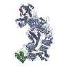
|
|---|---|
| 1 |
|
-Components
| #1: Protein | Mass: 19766.912 Da / Num. of mol.: 1 Source method: isolated from a genetically manipulated source Source: (gene. exp.) synthetic construct (others) / Production host:  |
|---|---|
| #2: Protein | Mass: 286462.781 Da / Num. of mol.: 1 Source method: isolated from a genetically manipulated source Source: (gene. exp.) Homo sapiens (human) / Gene: LRRK2, PARK8 / Production host:   Spodoptera frugiperda (fall armyworm) Spodoptera frugiperda (fall armyworm)References: UniProt: Q5S007, non-specific serine/threonine protein kinase, Hydrolases; Acting on acid anhydrides; Acting on GTP to facilitate cellular and subcellular movement |
| #3: Chemical | ChemComp-A1N / (  Mass: 379.456 Da / Num. of mol.: 1 / Source method: obtained synthetically / Formula: C21H25N5O2 / Feature type: SUBJECT OF INVESTIGATION Mass: 379.456 Da / Num. of mol.: 1 / Source method: obtained synthetically / Formula: C21H25N5O2 / Feature type: SUBJECT OF INVESTIGATION |
| #4: Chemical | ChemComp-GDP /   Type: RNA linking / Mass: 443.201 Da / Num. of mol.: 1 / Source method: obtained synthetically / Formula: C10H15N5O11P2 / Comment: GDP, energy-carrying molecule*YM Type: RNA linking / Mass: 443.201 Da / Num. of mol.: 1 / Source method: obtained synthetically / Formula: C10H15N5O11P2 / Comment: GDP, energy-carrying molecule*YM |
| Has ligand of interest | Y |
| Has protein modification | N |
-Experimental details
-Experiment
| Experiment | Method: ELECTRON MICROSCOPY |
|---|---|
| EM experiment | Aggregation state: PARTICLE / 3D reconstruction method: single particle reconstruction |
- Sample preparation
| Component | Name: Full-length LRRK2 (I2020T mutation) bound to MLi-2 / Type: COMPLEX / Entity ID: #1-#2 / Source: RECOMBINANT | |||||||||||||||||||||||||||||||||||
|---|---|---|---|---|---|---|---|---|---|---|---|---|---|---|---|---|---|---|---|---|---|---|---|---|---|---|---|---|---|---|---|---|---|---|---|---|
| Molecular weight | Value: 0.286 MDa / Experimental value: YES | |||||||||||||||||||||||||||||||||||
| Source (natural) | Organism: Homo sapiens (human) | |||||||||||||||||||||||||||||||||||
| Source (recombinant) | Organism: Spodoptera frugiperda (fall armyworm) | |||||||||||||||||||||||||||||||||||
| Buffer solution | pH: 7.4 | |||||||||||||||||||||||||||||||||||
| Buffer component |
| |||||||||||||||||||||||||||||||||||
| Specimen | Conc.: 1.43 mg/ml / Embedding applied: NO / Shadowing applied: NO / Staining applied: NO / Vitrification applied: YES | |||||||||||||||||||||||||||||||||||
| Specimen support | Grid material: GOLD / Grid mesh size: 200 divisions/in. / Grid type: UltrAuFoil R2/2 | |||||||||||||||||||||||||||||||||||
| Vitrification | Instrument: FEI VITROBOT MARK IV / Cryogen name: ETHANE / Humidity: 100 % / Chamber temperature: 277.15 K Details: Blot force = 3 Blot time = 4 seconds Wait time = 20 seconds |
- Electron microscopy imaging
| Experimental equipment |  Model: Titan Krios / Image courtesy: FEI Company |
|---|---|
| Microscopy | Model: FEI TITAN KRIOS |
| Electron gun | Electron source:  FIELD EMISSION GUN / Accelerating voltage: 300 kV / Illumination mode: FLOOD BEAM FIELD EMISSION GUN / Accelerating voltage: 300 kV / Illumination mode: FLOOD BEAM |
| Electron lens | Mode: BRIGHT FIELD / Nominal magnification: 130000 X / Nominal defocus max: 4000 nm / Nominal defocus min: 1000 nm / Cs: 2.7 mm / C2 aperture diameter: 50 µm |
| Specimen holder | Cryogen: NITROGEN / Specimen holder model: FEI TITAN KRIOS AUTOGRID HOLDER |
| Image recording | Average exposure time: 11 sec. / Electron dose: 55 e/Å2 / Film or detector model: FEI FALCON IV (4k x 4k) / Num. of grids imaged: 1 / Num. of real images: 10488 |
- Processing
| EM software |
| ||||||||||||||||||||||||||||||||||||
|---|---|---|---|---|---|---|---|---|---|---|---|---|---|---|---|---|---|---|---|---|---|---|---|---|---|---|---|---|---|---|---|---|---|---|---|---|---|
| CTF correction | Type: PHASE FLIPPING AND AMPLITUDE CORRECTION | ||||||||||||||||||||||||||||||||||||
| Particle selection | Num. of particles selected: 220313 | ||||||||||||||||||||||||||||||||||||
| Symmetry | Point symmetry: C1 (asymmetric) | ||||||||||||||||||||||||||||||||||||
| 3D reconstruction | Resolution: 3.9 Å / Resolution method: FSC 0.143 CUT-OFF / Num. of particles: 102649 / Symmetry type: POINT | ||||||||||||||||||||||||||||||||||||
| Atomic model building | Protocol: AB INITIO MODEL / Space: REAL | ||||||||||||||||||||||||||||||||||||
| Atomic model building | PDB-ID: 7LHW Pdb chain-ID: A / Accession code: 7LHW / Chain residue range: 1-2527 / Pdb chain residue range: 1-2527 / Source name: PDB / Type: experimental model | ||||||||||||||||||||||||||||||||||||
| Refine LS restraints |
|

