[English] 日本語
 Yorodumi
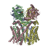
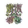
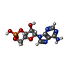
- PDB-8t50: Open human HCN1 F186C S264C bound to cAMP, reconstituted in LMNG + SPL -
+ Open data
- Basic information
| Entry | Database: PDB / ID: 8t50 | |||||||||
|---|---|---|---|---|---|---|---|---|---|---|
| Title | Open human HCN1 F186C S264C bound to cAMP, reconstituted in LMNG + SPL | |||||||||
 Components Components | Potassium/sodium hyperpolarization-activated cyclic nucleotide-gated channel 1 | |||||||||
 Keywords Keywords | TRANSPORT PROTEIN / Membrane Protein / Ion Channel | |||||||||
| Function / homology |  Function and homology information Function and homology information: / positive regulation of membrane hyperpolarization / HCN channels / general adaptation syndrome, behavioral process / HCN channel complex / retinal cone cell development / intracellularly cAMP-activated cation channel activity / negative regulation of action potential / regulation of SA node cell action potential / maternal behavior ...: / positive regulation of membrane hyperpolarization / HCN channels / general adaptation syndrome, behavioral process / HCN channel complex / retinal cone cell development / intracellularly cAMP-activated cation channel activity / negative regulation of action potential / regulation of SA node cell action potential / maternal behavior / apical dendrite / sodium ion import across plasma membrane / response to L-glutamate / apical protein localization / negative regulation of synaptic transmission, glutamatergic / voltage-gated sodium channel activity / voltage-gated monoatomic cation channel activity / regulation of membrane depolarization / potassium ion import across plasma membrane / phosphatidylinositol-3,4,5-trisphosphate binding / regulation of heart rate by cardiac conduction / voltage-gated potassium channel activity / potassium channel activity / cAMP binding / neuronal action potential / cellular response to interferon-beta / phosphatidylinositol-4,5-bisphosphate binding / potassium ion transmembrane transport / presynaptic active zone membrane / dendrite membrane / axon terminus / cellular response to cAMP / sodium ion transmembrane transport / dendritic shaft / regulation of membrane potential / response to calcium ion / basolateral plasma membrane / protein homotetramerization / postsynaptic membrane / axon / neuronal cell body / dendrite / glutamatergic synapse / cell surface / identical protein binding / plasma membrane Similarity search - Function | |||||||||
| Biological species |  Homo sapiens (human) Homo sapiens (human) | |||||||||
| Method | ELECTRON MICROSCOPY / single particle reconstruction / cryo EM / Resolution: 3.6 Å | |||||||||
 Authors Authors | Burtscher, V. / Mount, J. / Cowgill, J. / Chang, Y. / Bickel, K. / Yuan, P. / Chanda, B. | |||||||||
| Funding support |  United States, 2items United States, 2items
| |||||||||
 Citation Citation |  Journal: Nat Commun / Year: 2024 Journal: Nat Commun / Year: 2024Title: Structural basis for hyperpolarization-dependent opening of human HCN1 channel. Authors: Verena Burtscher / Jonathan Mount / Jian Huang / John Cowgill / Yongchang Chang / Kathleen Bickel / Jianhan Chen / Peng Yuan / Baron Chanda /   Abstract: Hyperpolarization and cyclic nucleotide (HCN) activated ion channels are critical for the automaticity of action potentials in pacemaking and rhythmic electrical circuits in the human body. Unlike ...Hyperpolarization and cyclic nucleotide (HCN) activated ion channels are critical for the automaticity of action potentials in pacemaking and rhythmic electrical circuits in the human body. Unlike most voltage-gated ion channels, the HCN and related plant ion channels activate upon membrane hyperpolarization. Although functional studies have identified residues in the interface between the voltage-sensing and pore domain as crucial for inverted electromechanical coupling, the structural mechanisms for this unusual voltage-dependence remain unclear. Here, we present cryo-electron microscopy structures of human HCN1 corresponding to Closed, Open, and a putative Intermediate state. Our structures reveal that the downward motion of the gating charges past the charge transfer center is accompanied by concomitant unwinding of the inner end of the S4 and S5 helices, disrupting the tight gating interface observed in the Closed state structure. This helix-coil transition at the intracellular gating interface accompanies a concerted iris-like dilation of the pore helices and underlies the reversed voltage dependence of HCN channels. | |||||||||
| History |
|
- Structure visualization
| Structure viewer | Molecule:  Molmil Molmil Jmol/JSmol Jmol/JSmol |
|---|
- Downloads & links
- Download
| PDBx/mmCIF format |  8t50.cif.gz 8t50.cif.gz | 338.2 KB | Display |  PDBx/mmCIF format PDBx/mmCIF format |
|---|---|---|---|---|
| PDB format |  pdb8t50.ent.gz pdb8t50.ent.gz | 241.6 KB | Display |  PDB format PDB format |
| PDBx/mmJSON format |  8t50.json.gz 8t50.json.gz | Tree view |  PDBx/mmJSON format PDBx/mmJSON format | |
| Others |  Other downloads Other downloads |
-Validation report
| Arichive directory |  https://data.pdbj.org/pub/pdb/validation_reports/t5/8t50 https://data.pdbj.org/pub/pdb/validation_reports/t5/8t50 ftp://data.pdbj.org/pub/pdb/validation_reports/t5/8t50 ftp://data.pdbj.org/pub/pdb/validation_reports/t5/8t50 | HTTPS FTP |
|---|
-Related structure data
| Related structure data |  41041MC  8t4mC  8t4yC C: citing same article ( M: map data used to model this data |
|---|---|
| Similar structure data | Similarity search - Function & homology  F&H Search F&H Search |
- Links
Links
- Assembly
| Deposited unit | 
|
|---|---|
| 1 |
|
- Components
| #1: Protein | Mass: 98871.820 Da / Num. of mol.: 4 / Mutation: F186C, S264C Source method: isolated from a genetically manipulated source Source: (gene. exp.)  Homo sapiens (human) / Gene: HCN1, BCNG1 / Production host: Homo sapiens (human) / Gene: HCN1, BCNG1 / Production host:  Homo sapiens (human) / References: UniProt: O60741 Homo sapiens (human) / References: UniProt: O60741#2: Chemical | ChemComp-CMP / Has ligand of interest | Y | Has protein modification | Y | |
|---|
-Experimental details
-Experiment
| Experiment | Method: ELECTRON MICROSCOPY |
|---|---|
| EM experiment | Aggregation state: PARTICLE / 3D reconstruction method: single particle reconstruction |
- Sample preparation
| Component | Name: hHCN1 F186C S264C / Type: COMPLEX / Details: Homomeric Tetramer / Entity ID: #1 / Source: RECOMBINANT | |||||||||||||||||||||||||||||||||||||||||||||
|---|---|---|---|---|---|---|---|---|---|---|---|---|---|---|---|---|---|---|---|---|---|---|---|---|---|---|---|---|---|---|---|---|---|---|---|---|---|---|---|---|---|---|---|---|---|---|
| Source (natural) | Organism:  Homo sapiens (human) Homo sapiens (human) | |||||||||||||||||||||||||||||||||||||||||||||
| Source (recombinant) | Organism:  Homo sapiens (human) Homo sapiens (human) | |||||||||||||||||||||||||||||||||||||||||||||
| Buffer solution | pH: 8 | |||||||||||||||||||||||||||||||||||||||||||||
| Buffer component |
| |||||||||||||||||||||||||||||||||||||||||||||
| Specimen | Conc.: 2 mg/ml / Embedding applied: NO / Shadowing applied: NO / Staining applied: NO / Vitrification applied: YES | |||||||||||||||||||||||||||||||||||||||||||||
| Specimen support | Grid material: COPPER / Grid mesh size: 200 divisions/in. / Grid type: Quantifoil R2/2 | |||||||||||||||||||||||||||||||||||||||||||||
| Vitrification | Instrument: FEI VITROBOT MARK IV / Cryogen name: ETHANE |
- Electron microscopy imaging
| Experimental equipment |  Model: Titan Krios / Image courtesy: FEI Company |
|---|---|
| Microscopy | Model: FEI TITAN KRIOS |
| Electron gun | Electron source:  FIELD EMISSION GUN / Accelerating voltage: 300 kV / Illumination mode: FLOOD BEAM FIELD EMISSION GUN / Accelerating voltage: 300 kV / Illumination mode: FLOOD BEAM |
| Electron lens | Mode: BRIGHT FIELD / Nominal defocus max: 2400 nm / Nominal defocus min: 800 nm |
| Image recording | Electron dose: 54.25 e/Å2 / Film or detector model: FEI FALCON IV (4k x 4k) / Num. of grids imaged: 2 |
- Processing
| EM software |
| ||||||||||||||||||||||||||||
|---|---|---|---|---|---|---|---|---|---|---|---|---|---|---|---|---|---|---|---|---|---|---|---|---|---|---|---|---|---|
| CTF correction | Type: PHASE FLIPPING AND AMPLITUDE CORRECTION | ||||||||||||||||||||||||||||
| Particle selection | Num. of particles selected: 867853 | ||||||||||||||||||||||||||||
| Symmetry | Point symmetry: C4 (4 fold cyclic) | ||||||||||||||||||||||||||||
| 3D reconstruction | Resolution: 3.6 Å / Resolution method: FSC 0.143 CUT-OFF / Num. of particles: 438138 / Num. of class averages: 3 / Symmetry type: POINT | ||||||||||||||||||||||||||||
| Atomic model building | Protocol: FLEXIBLE FIT / Space: REAL | ||||||||||||||||||||||||||||
| Atomic model building | PDB-ID: 6UQF Accession code: 6UQF / Source name: PDB / Type: experimental model | ||||||||||||||||||||||||||||
| Refine LS restraints |
|
 Movie
Movie Controller


 PDBj
PDBj


