 Movie
Movie Controller

[English] 日本語
 Yorodumi
- PDB-8t0z: Human liver-type glutaminase (K253A) with L-Gln, filamentous form -
+ Open data

- Basic information
| Entry | Database: PDB / ID: 8t0z | ||||||||||||||||||||||||
|---|---|---|---|---|---|---|---|---|---|---|---|---|---|---|---|---|---|---|---|---|---|---|---|---|---|
| Title | Human liver-type glutaminase (K253A) with L-Gln, filamentous form | ||||||||||||||||||||||||
 Components Components | Glutaminase liver isoform, mitochondrial | ||||||||||||||||||||||||
 Keywords Keywords | HYDROLASE / Metabolic / Cancer | ||||||||||||||||||||||||
| Function / homology |  Function and homology information Function and homology informationL-glutamine catabolic process / glutaminase / Glutamate and glutamine metabolism / Glutamate Neurotransmitter Release Cycle / : / glutaminase activity / amino acid metabolic process / reactive oxygen species metabolic process / TP53 Regulates Metabolic Genes / regulation of apoptotic process ...L-glutamine catabolic process / glutaminase / Glutamate and glutamine metabolism / Glutamate Neurotransmitter Release Cycle / : / glutaminase activity / amino acid metabolic process / reactive oxygen species metabolic process / TP53 Regulates Metabolic Genes / regulation of apoptotic process / mitochondrial matrix / mitochondrion Similarity search - Function | ||||||||||||||||||||||||
| Biological species |  Homo sapiens (human) Homo sapiens (human) | ||||||||||||||||||||||||
| Method | ELECTRON MICROSCOPY / helical reconstruction / cryo EM / Resolution: 3.3 Å | ||||||||||||||||||||||||
 Authors Authors | Feng, S. / Aplin, C. / Nguyen, T.-T.T. / Milano, S.K. / Cerione, R.A. | ||||||||||||||||||||||||
| Funding support |  United States, 7items United States, 7items
| ||||||||||||||||||||||||
 Citation Citation | Journal: Nat Commun / Year: 2024 Title: Filament formation drives catalysis by glutaminase enzymes important in cancer progression. Authors: Shi Feng / Cody Aplin / Thuy-Tien T Nguyen / Shawn K Milano / Richard A Cerione / Abstract: The glutaminase enzymes GAC and GLS2 catalyze the hydrolysis of glutamine to glutamate, satisfying the 'glutamine addiction' of cancer cells. They are the targets of anti-cancer drugs; however, their ...The glutaminase enzymes GAC and GLS2 catalyze the hydrolysis of glutamine to glutamate, satisfying the 'glutamine addiction' of cancer cells. They are the targets of anti-cancer drugs; however, their mechanisms of activation and catalytic activity have been unclear. Here we demonstrate that the ability of GAC and GLS2 to form filaments is directly coupled to their catalytic activity and present their cryo-EM structures which provide a view of the conformational states essential for catalysis. Filament formation guides an 'activation loop' to assume a specific conformation that works together with a 'lid' to close over the active site and position glutamine for nucleophilic attack by an essential serine. Our findings highlight how ankyrin repeats on GLS2 regulate enzymatic activity, while allosteric activators stabilize, and clinically relevant inhibitors block, filament formation that enables glutaminases to catalyze glutaminolysis and support cancer progression. | ||||||||||||||||||||||||
| History |
|
- Structure visualization
| Structure viewer | Molecule: MolmilJmol/JSmol |
|---|
- Downloads & links
-Download
| PDBx/mmCIF format | 8t0z.cif.gz | 1.7 MB | Display | PDBx/mmCIF format |
|---|---|---|---|---|
| PDB format | pdb8t0z.ent.gz | 1.5 MB | Display | PDB format |
| PDBx/mmJSON format | 8t0z.json.gz | Tree view | PDBx/mmJSON format | |
| Others |  Other downloads Other downloads |
-Validation report
| Arichive directory | https://data.pdbj.org/pub/pdb/validation_reports/t0/8t0zftp://data.pdbj.org/pub/pdb/validation_reports/t0/8t0z | HTTPS FTP |
|---|
-Related structure data
| Related structure data |  40950MC 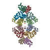 8szjC  8szlC M: map data used to model this data C: citing same article ( |
|---|---|
| Similar structure data |


-Links
 PDBj
PDBj

- Assembly
| Deposited unit | 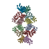
|
|---|---|
| 1 |
|
-Components
| #1: Protein | Mass: 66351.617 Da / Num. of mol.: 12 / Mutation: K253A Source method: isolated from a genetically manipulated source Source: (gene. exp.) Homo sapiens (human) / Gene: GLS2, GA / Production host:  #2: Chemical | ChemComp-GLN /   Type: L-peptide linking / Mass: 146.144 Da / Num. of mol.: 12 / Source method: obtained synthetically / Formula: C5H10N2O3 / Feature type: SUBJECT OF INVESTIGATION Type: L-peptide linking / Mass: 146.144 Da / Num. of mol.: 12 / Source method: obtained synthetically / Formula: C5H10N2O3 / Feature type: SUBJECT OF INVESTIGATIONHas ligand of interest | Y | |
|---|
-Experimental details
-Experiment
| Experiment | Method: ELECTRON MICROSCOPY |
|---|---|
| EM experiment | Aggregation state: FILAMENT / 3D reconstruction method: helical reconstruction |
- Sample preparation
| Component | Name: Human liver-type glutaminase (K253A) with L-Gln, filamentous form Type: COMPLEX / Entity ID: #1 / Source: RECOMBINANT |
|---|---|
| Molecular weight | Experimental value: NO |
| Source (natural) | Organism: Homo sapiens (human) |
| Source (recombinant) | Organism: |
| Buffer solution | pH: 8 |
| Specimen | Embedding applied: NO / Shadowing applied: NO / Staining applied: NO / Vitrification applied: YES |
| Vitrification | Cryogen name: ETHANE-PROPANE |
- Electron microscopy imaging
| Experimental equipment |  Model: Titan Krios / Image courtesy: FEI Company |
|---|---|
| Microscopy | Model: FEI TITAN KRIOS |
| Electron gun | Electron source:  FIELD EMISSION GUN / Accelerating voltage: 300 kV / Illumination mode: FLOOD BEAM FIELD EMISSION GUN / Accelerating voltage: 300 kV / Illumination mode: FLOOD BEAM |
| Electron lens | Mode: BRIGHT FIELD / Nominal defocus max: 2500 nm / Nominal defocus min: 800 nm |
| Image recording | Electron dose: 50 e/Å2 / Film or detector model: GATAN K3 BIOQUANTUM (6k x 4k) |
- Processing
| EM software | Name: PHENIX / Version: 1.20.1_4487: / Category: model refinement |
|---|---|
| CTF correction | Type: NONE |
| Helical symmerty | Angular rotation/subunit: 48 ° / Axial rise/subunit: 66 Å / Axial symmetry: C1 |
| 3D reconstruction | Resolution: 3.3 Å / Resolution method: FSC 0.143 CUT-OFF / Num. of particles: 48000 / Symmetry type: HELICAL |
