 Movie
Movie Controller


+ Open data

- Basic information
| Entry | Database: PDB / ID: 8t06 | ||||||
|---|---|---|---|---|---|---|---|
| Title | Structure of mouse Myomaker mutant-R107A bound to Fab18G7 | ||||||
 Components Components |
| ||||||
 Keywords Keywords | MEMBRANE PROTEIN | ||||||
| Function / homology |  Function and homology information Function and homology informationpositive regulation of skeletal muscle hypertrophy / : / myoblast fusion involved in skeletal muscle regeneration / skeletal muscle tissue regeneration / myoblast fusion / muscle organ development / Golgi membrane / plasma membrane Similarity search - Function | ||||||
| Biological species |  | ||||||
| Method | ELECTRON MICROSCOPY / single particle reconstruction / cryo EM / Resolution: 3.32 Å | ||||||
 Authors Authors | Long, T. / Li, X. | ||||||
| Funding support |  United States, 1items United States, 1items
| ||||||
 Citation Citation | Journal: Nat Struct Mol Biol / Year: 2023 Title: Cryo-EM structures of Myomaker reveal a molecular basis for myoblast fusion. Authors: Tao Long / Yichi Zhang / Linda Donnelly / Hui Li / Yu-Chung Pien / Ning Liu / Eric N Olson / Xiaochun Li / Abstract: The fusion of mononucleated myoblasts produces multinucleated muscle fibers leading to the formation of skeletal muscle. Myomaker, a skeletal muscle-specific membrane protein, is essential for ...The fusion of mononucleated myoblasts produces multinucleated muscle fibers leading to the formation of skeletal muscle. Myomaker, a skeletal muscle-specific membrane protein, is essential for myoblast fusion. Here we report the cryo-EM structures of mouse Myomaker (mMymk) and Ciona robusta Myomaker (cMymk). Myomaker contains seven transmembrane helices (TMs) that adopt a G-protein-coupled receptor-like fold. TMs 2-4 form a dimeric interface, while TMs 3 and 5-7 create a lipid-binding site that holds the polar head of a phospholipid and allows the alkyl tails to insert into Myomaker. The similarity of cMymk and mMymk suggests a conserved Myomaker-mediated cell fusion mechanism across evolutionarily distant species. Functional analyses demonstrate the essentiality of the dimeric interface and the lipid-binding site for fusogenic activity, and heterologous cell-cell fusion assays show the importance of transcellular interactions of Myomaker protomers for myoblast fusion. Together, our findings provide structural and functional insights into the process of myoblast fusion. | ||||||
| History |
|
- Structure visualization
| Structure viewer | Molecule: MolmilJmol/JSmol |
|---|
- Downloads & links
-Download
| PDBx/mmCIF format | 8t06.cif.gz | 174.8 KB | Display | PDBx/mmCIF format |
|---|---|---|---|---|
| PDB format | pdb8t06.ent.gz | 136.8 KB | Display | PDB format |
| PDBx/mmJSON format | 8t06.json.gz | Tree view | PDBx/mmJSON format | |
| Others |  Other downloads Other downloads |
-Validation report
| Arichive directory | https://data.pdbj.org/pub/pdb/validation_reports/t0/8t06ftp://data.pdbj.org/pub/pdb/validation_reports/t0/8t06 | HTTPS FTP |
|---|
-Related structure data
| Related structure data |  40936MC 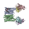 8t03C 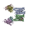 8t04C  8t05C 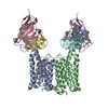 8t07C M: map data used to model this data C: citing same article ( |
|---|---|
| Similar structure data |


-Links
 PDBj
PDBj

- Assembly
| Deposited unit | 
|
|---|---|
| 1 |
|
-Components
| #1: Protein | Mass: 24731.354 Da / Num. of mol.: 2 / Mutation: R107A Source method: isolated from a genetically manipulated source Source: (gene. exp.)   Spodoptera frugiperda (fall armyworm) / References: UniProt: Q9D1N4 Spodoptera frugiperda (fall armyworm) / References: UniProt: Q9D1N4#2: Antibody | Mass: 13318.994 Da / Num. of mol.: 2 Source method: isolated from a genetically manipulated source Source: (gene. exp.)  Homo sapiens (human) Homo sapiens (human)#3: Antibody | Mass: 11809.201 Da / Num. of mol.: 2 Source method: isolated from a genetically manipulated source Source: (gene. exp.) Homo sapiens (human)#4: Chemical |   Mass: 65.409 Da / Num. of mol.: 2 / Source method: obtained synthetically / Formula: Zn Mass: 65.409 Da / Num. of mol.: 2 / Source method: obtained synthetically / Formula: ZnHas ligand of interest | N | Has protein modification | Y | |
|---|
-Experimental details
-Experiment
| Experiment | Method: ELECTRON MICROSCOPY |
|---|---|
| EM experiment | Aggregation state: PARTICLE / 3D reconstruction method: single particle reconstruction |
- Sample preparation
| Component |
| ||||||||||||||||||||||||
|---|---|---|---|---|---|---|---|---|---|---|---|---|---|---|---|---|---|---|---|---|---|---|---|---|---|
| Source (natural) |
| ||||||||||||||||||||||||
| Source (recombinant) |
| ||||||||||||||||||||||||
| Buffer solution | pH: 7.5 | ||||||||||||||||||||||||
| Specimen | Embedding applied: NO / Shadowing applied: NO / Staining applied: NO / Vitrification applied: YES | ||||||||||||||||||||||||
| Vitrification | Cryogen name: ETHANE |
- Electron microscopy imaging
| Experimental equipment |  Model: Titan Krios / Image courtesy: FEI Company |
|---|---|
| Microscopy | Model: FEI TITAN KRIOS |
| Electron gun | Electron source:  FIELD EMISSION GUN / Accelerating voltage: 300 kV / Illumination mode: FLOOD BEAM FIELD EMISSION GUN / Accelerating voltage: 300 kV / Illumination mode: FLOOD BEAM |
| Electron lens | Mode: BRIGHT FIELD / Nominal defocus max: 1800 nm / Nominal defocus min: 800 nm |
| Image recording | Electron dose: 60 e/Å2 / Film or detector model: GATAN K3 (6k x 4k) |
- Processing
| EM software | Name: REFMAC / Version: 5.8.0267 / Category: model refinement | ||||||||||||||||||||||||||||||||||||||||||||||||||||||||||||||||||||||||||||||||||||||||||||||||||||||||||
|---|---|---|---|---|---|---|---|---|---|---|---|---|---|---|---|---|---|---|---|---|---|---|---|---|---|---|---|---|---|---|---|---|---|---|---|---|---|---|---|---|---|---|---|---|---|---|---|---|---|---|---|---|---|---|---|---|---|---|---|---|---|---|---|---|---|---|---|---|---|---|---|---|---|---|---|---|---|---|---|---|---|---|---|---|---|---|---|---|---|---|---|---|---|---|---|---|---|---|---|---|---|---|---|---|---|---|---|
| CTF correction | Type: NONE | ||||||||||||||||||||||||||||||||||||||||||||||||||||||||||||||||||||||||||||||||||||||||||||||||||||||||||
| 3D reconstruction | Resolution: 3.32 Å / Resolution method: FSC 0.143 CUT-OFF / Num. of particles: 154081 / Symmetry type: POINT | ||||||||||||||||||||||||||||||||||||||||||||||||||||||||||||||||||||||||||||||||||||||||||||||||||||||||||
| Refinement | Resolution: 3.32→249 Å / Cor.coef. Fo:Fc: 0.82 / Cor.coef. Fo:Fc free: 0.863 / SU B: 11.715 / SU ML: 0.166 / Cross valid method: THROUGHOUT / ESU R: 0.12 / ESU R Free: 0.114 / Stereochemistry target values: MAXIMUM LIKELIHOOD / Details: HYDROGENS HAVE BEEN ADDED IN THE RIDING POSITIONS
| ||||||||||||||||||||||||||||||||||||||||||||||||||||||||||||||||||||||||||||||||||||||||||||||||||||||||||
| Solvent computation | Ion probe radii: 0.8 Å / Shrinkage radii: 0.8 Å / VDW probe radii: 1.2 Å / Solvent model: MASK | ||||||||||||||||||||||||||||||||||||||||||||||||||||||||||||||||||||||||||||||||||||||||||||||||||||||||||
| Displacement parameters | Biso mean: 139.059 Å2
| ||||||||||||||||||||||||||||||||||||||||||||||||||||||||||||||||||||||||||||||||||||||||||||||||||||||||||
| Refinement step | Cycle: 1 / Total: 6712 | ||||||||||||||||||||||||||||||||||||||||||||||||||||||||||||||||||||||||||||||||||||||||||||||||||||||||||
| Refine LS restraints |
|