 Movie
Movie Controller


+ Open data

- Basic information
| Entry |  | |||||||||
|---|---|---|---|---|---|---|---|---|---|---|
| Title | Structure of mouse Myomaker mutant-Y118A bound to Fab18G7 | |||||||||
 Map data Map data | ||||||||||
 Sample Sample |
| |||||||||
 Keywords Keywords | MEMBRANE PROTEIN | |||||||||
| Function / homology |  Function and homology information Function and homology informationpositive regulation of skeletal muscle hypertrophy / : / myoblast fusion involved in skeletal muscle regeneration / skeletal muscle tissue regeneration / myoblast fusion / muscle organ development / Golgi membrane / plasma membrane Similarity search - Function | |||||||||
| Biological species |  | |||||||||
| Method | single particle reconstruction / cryo EM / Resolution: 3.38 Å | |||||||||
 Authors Authors | Long T / Li X | |||||||||
| Funding support |  United States, 1 items United States, 1 items
| |||||||||
 Citation Citation | Journal: Nat Struct Mol Biol / Year: 2023 Title: Cryo-EM structures of Myomaker reveal a molecular basis for myoblast fusion. Authors: Tao Long / Yichi Zhang / Linda Donnelly / Hui Li / Yu-Chung Pien / Ning Liu / Eric N Olson / Xiaochun Li / Abstract: The fusion of mononucleated myoblasts produces multinucleated muscle fibers leading to the formation of skeletal muscle. Myomaker, a skeletal muscle-specific membrane protein, is essential for ...The fusion of mononucleated myoblasts produces multinucleated muscle fibers leading to the formation of skeletal muscle. Myomaker, a skeletal muscle-specific membrane protein, is essential for myoblast fusion. Here we report the cryo-EM structures of mouse Myomaker (mMymk) and Ciona robusta Myomaker (cMymk). Myomaker contains seven transmembrane helices (TMs) that adopt a G-protein-coupled receptor-like fold. TMs 2-4 form a dimeric interface, while TMs 3 and 5-7 create a lipid-binding site that holds the polar head of a phospholipid and allows the alkyl tails to insert into Myomaker. The similarity of cMymk and mMymk suggests a conserved Myomaker-mediated cell fusion mechanism across evolutionarily distant species. Functional analyses demonstrate the essentiality of the dimeric interface and the lipid-binding site for fusogenic activity, and heterologous cell-cell fusion assays show the importance of transcellular interactions of Myomaker protomers for myoblast fusion. Together, our findings provide structural and functional insights into the process of myoblast fusion. | |||||||||
| History |
|
- Structure visualization
| Supplemental images |
|---|

- Downloads & links
-EMDB archive
| Map data | emd_40937.map.gz | 97.1 MB | EMDB map data format | |
|---|---|---|---|---|
| Header (meta data) | emd-40937-v30.xmlemd-40937.xml | 16.7 KB 16.7 KB | Display Display | EMDB header |
| Images | 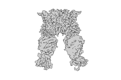 emd_40937.png emd_40937.png | 49.4 KB | ||
| Masks | emd_40937_msk_1.map | 103 MB | Mask map | |
| Filedesc metadata | emd-40937.cif.gz | 5.6 KB | ||
| Others | emd_40937_half_map_1.map.gzemd_40937_half_map_2.map.gz | 95.3 MB 95.3 MB | ||
| Archive directory |  http://ftp.pdbj.org/pub/emdb/structures/EMD-40937ftp://ftp.pdbj.org/pub/emdb/structures/EMD-40937 http://ftp.pdbj.org/pub/emdb/structures/EMD-40937ftp://ftp.pdbj.org/pub/emdb/structures/EMD-40937 | HTTPS FTP |
-Related structure data
| Related structure data | 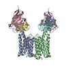 8t07MC 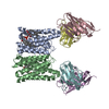 8t03C 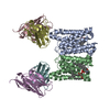 8t04C  8t05C 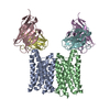 8t06C M: atomic model generated by this map C: citing same article ( |
|---|---|
| Similar structure data |


-Links
| EMDB pages | EMDB (EBI/PDBe) / EMDataResource |
|---|
-Map
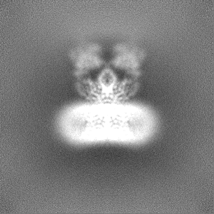
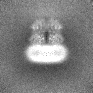
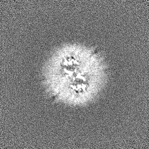
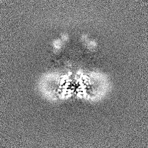
| File | Download / File: emd_40937.map.gz / Format: CCP4 / Size: 103 MB / Type: IMAGE STORED AS FLOATING POINT NUMBER (4 BYTES) | ||||||||||||||||||||||||||||||||||||
|---|---|---|---|---|---|---|---|---|---|---|---|---|---|---|---|---|---|---|---|---|---|---|---|---|---|---|---|---|---|---|---|---|---|---|---|---|---|
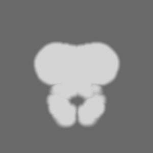
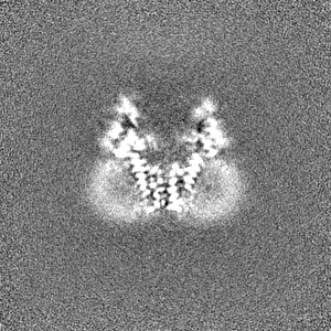
| Projections & slices | Image control
Images are generated by Spider. | ||||||||||||||||||||||||||||||||||||
| Voxel size | X=Y=Z: 0.83 Å | ||||||||||||||||||||||||||||||||||||
| Density |
| ||||||||||||||||||||||||||||||||||||
| Symmetry | Space group: 1 | ||||||||||||||||||||||||||||||||||||
| Details | EMDB XML:
|
 Z (Sec.)
 Y (Row.)
 X (Col.)


-Supplemental data
-Mask #1
| File | emd_40937_msk_1.map | ||||||||||||
|---|---|---|---|---|---|---|---|---|---|---|---|---|---|
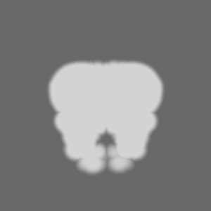
| Projections & Slices |
| ||||||||||||
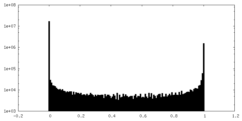
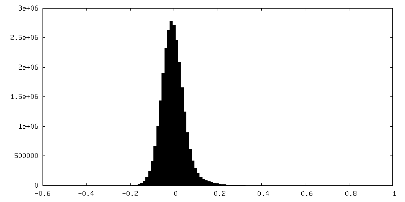
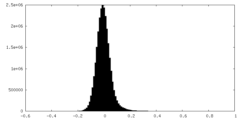
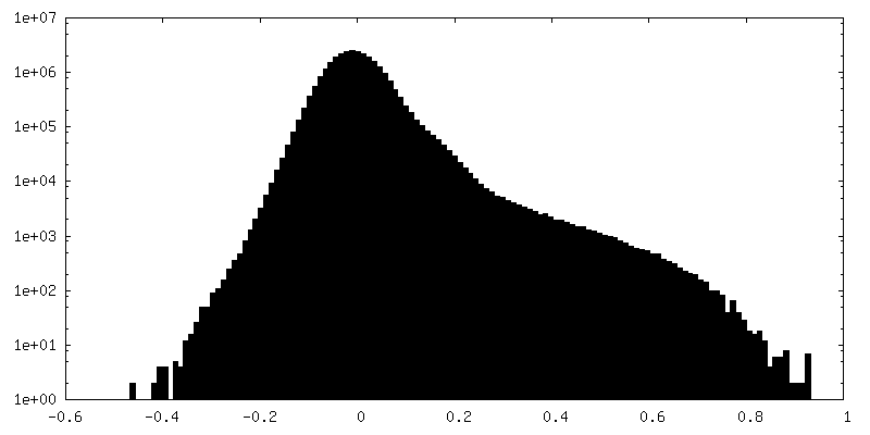
| Density Histograms |


-Half map: #2
| File | emd_40937_half_map_1.map | ||||||||||||
|---|---|---|---|---|---|---|---|---|---|---|---|---|---|
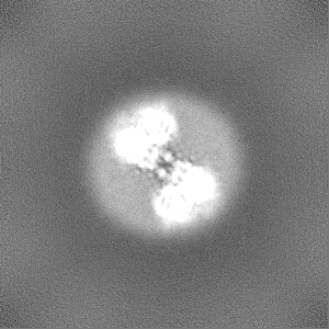
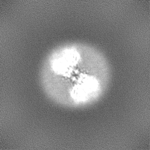
| Projections & Slices |
| ||||||||||||
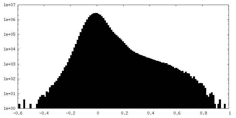
| Density Histograms |
-Half map: #1
| File | emd_40937_half_map_2.map | ||||||||||||
|---|---|---|---|---|---|---|---|---|---|---|---|---|---|
| Projections & Slices |
| ||||||||||||
| Density Histograms |

- Sample components
-Entire : Complex of mouse Myomaker mutant-Y118A with Fab18G7
| Entire | Name: Complex of mouse Myomaker mutant-Y118A with Fab18G7 |
|---|---|
| Components |
|
-Supramolecule #1: Complex of mouse Myomaker mutant-Y118A with Fab18G7
| Supramolecule | Name: Complex of mouse Myomaker mutant-Y118A with Fab18G7 / type: complex / ID: 1 / Parent: 0 / Macromolecule list: #1-#3 |
|---|
-Supramolecule #2: mouse Myomaker mutant-Y118A
| Supramolecule | Name: mouse Myomaker mutant-Y118A / type: complex / ID: 2 / Parent: 1 / Macromolecule list: #1 |
|---|---|
| Source (natural) | Organism: |
-Supramolecule #3: Fab18G7
| Supramolecule | Name: Fab18G7 / type: complex / ID: 3 / Parent: 1 / Macromolecule list: #2-#3 |
|---|---|
| Source (natural) | Organism: |
-Macromolecule #1: Protein myomaker
| Macromolecule | Name: Protein myomaker / type: protein_or_peptide / ID: 1 / Number of copies: 2 / Enantiomer: LEVO |
|---|---|
| Source (natural) | Organism: |
| Molecular weight | Theoretical: 24.725371 KDa |
| Recombinant expression | Organism:   Spodoptera frugiperda (fall armyworm) Spodoptera frugiperda (fall armyworm) |
| Sequence | String: MGTVVAKLLL PTLSSLAFLP TVSIATKRRF YMEAMVYLFT MFFVAFSHAC DGPGLSVLCF MRRDILEYFS IYGTALSMWV SLMALADFD EPQRSTFTML GVLTIAVRTF HDRWGYGVAS GPIGTATLII AVKWLKKMKE KKGLYPDKSI YTQQIGPGLC F GALALMLR ...String: MGTVVAKLLL PTLSSLAFLP TVSIATKRRF YMEAMVYLFT MFFVAFSHAC DGPGLSVLCF MRRDILEYFS IYGTALSMWV SLMALADFD EPQRSTFTML GVLTIAVRTF HDRWGYGVAS GPIGTATLII AVKWLKKMKE KKGLYPDKSI YTQQIGPGLC F GALALMLR FFFEEWDYTY VHSFYHCALA MSFVLLLPKV NKKAGNAGAP AKLTFSTLCC TCV UniProtKB: Protein myomaker |
-Macromolecule #2: 18G7 Fab heavy chain
| Macromolecule | Name: 18G7 Fab heavy chain / type: protein_or_peptide / ID: 2 / Number of copies: 2 / Enantiomer: LEVO |
|---|---|
| Source (natural) | Organism: |
| Molecular weight | Theoretical: 13.318994 KDa |
| Recombinant expression | Organism:  Homo sapiens (human) Homo sapiens (human) |
| Sequence | String: QVTLKESGPG ILQPSQTLSL TCSFSGFSLS TSGMGVSWIR KPSGKGLEWL AHIFWDDDKR YNPSLKSRLT ISKDTSSNQV FLMITSIDT ADTATYYCAR RTWLLHAMDY WGQGTSVTVS S |
-Macromolecule #3: 18G7 Fab light chain
| Macromolecule | Name: 18G7 Fab light chain / type: protein_or_peptide / ID: 3 / Number of copies: 2 / Enantiomer: LEVO |
|---|---|
| Source (natural) | Organism: |
| Molecular weight | Theoretical: 11.809201 KDa |
| Recombinant expression | Organism: Homo sapiens (human) |
| Sequence | String: DIQMTQSPSS LSASLGGKVT ITCKASQDIN EYIAWYQHKP GKGPRLLIHY TSTLQPGIPS RFSGSGSGRD YSFSISNLEP EDIATYYCL QYDNLLWTFG GGTKLEIK |
-Macromolecule #4: ZINC ION
| Macromolecule | Name: ZINC ION / type: ligand / ID: 4 / Number of copies: 2 / Formula: ZN |
|---|---|
| Molecular weight | Theoretical: 65.409 Da |
-Experimental details
-Structure determination
| Method | cryo EM |
|---|---|
 Processing Processing | single particle reconstruction |
| Aggregation state | particle |
-Sample preparation
| Buffer | pH: 7.5 |
|---|---|
| Vitrification | Cryogen name: ETHANE |
- Electron microscopy
| Microscope | FEI TITAN KRIOS |
|---|---|
| Image recording | Film or detector model: GATAN K3 (6k x 4k) / Average electron dose: 60.0 e/Å2 |
| Electron beam | Acceleration voltage: 300 kV / Electron source:  FIELD EMISSION GUN FIELD EMISSION GUN |
| Electron optics | Illumination mode: FLOOD BEAM / Imaging mode: BRIGHT FIELD / Nominal defocus max: 1.8 µm / Nominal defocus min: 0.8 µm |
| Experimental equipment |  Model: Titan Krios / Image courtesy: FEI Company |
-Image processing
| Startup model | Type of model: OTHER |
|---|---|
| Final reconstruction | Resolution.type: BY AUTHOR / Resolution: 3.38 Å / Resolution method: FSC 0.143 CUT-OFF / Number images used: 124683 |
| Initial angle assignment | Type: NOT APPLICABLE |
| Final angle assignment | Type: NOT APPLICABLE |
