 Movie
Movie Controller

[English] 日本語
 Yorodumi
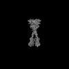
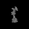
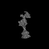
- PDB-8szi: Cryo-EM structure of PAM-free human calcium-sensing receptor CaSR... -
+ Open data

- Basic information
| Entry | Database: PDB / ID: 8szi | ||||||
|---|---|---|---|---|---|---|---|
| Title | Cryo-EM structure of PAM-free human calcium-sensing receptor CaSR-Gi complex in lipid nanodiscs | ||||||
 Components Components |
| ||||||
 Keywords Keywords | SIGNALING PROTEIN / Family C GPCR / Calcium-sensing Receptor (CaSR) / Heterotrimeric G protein / Cryo-EM / Lipid Nanodiscs / Positive Allosteric Modulator / Membrane Protein | ||||||
| Function / homology |  Function and homology information Function and homology informationregulation of presynaptic membrane potential / bile acid secretion / chemosensory behavior / response to fibroblast growth factor / cellular response to peptide / cellular response to vitamin D / negative regulation of adenylate cyclase activity / positive regulation of positive chemotaxis / Class C/3 (Metabotropic glutamate/pheromone receptors) / GTP metabolic process ...regulation of presynaptic membrane potential / bile acid secretion / chemosensory behavior / response to fibroblast growth factor / cellular response to peptide / cellular response to vitamin D / negative regulation of adenylate cyclase activity / positive regulation of positive chemotaxis / Class C/3 (Metabotropic glutamate/pheromone receptors) / GTP metabolic process / fat pad development / cellular response to hepatocyte growth factor stimulus / amino acid binding / branching morphogenesis of an epithelial tube / positive regulation of calcium ion import / regulation of calcium ion transport / positive regulation of NLRP3 inflammasome complex assembly / positive regulation of macroautophagy / positive regulation of vasoconstriction / anatomical structure morphogenesis / cellular response to low-density lipoprotein particle stimulus / detection of calcium ion / JNK cascade / Adenylate cyclase inhibitory pathway / ossification / axon terminus / response to ischemia / chloride transmembrane transport / cellular response to glucose stimulus / G protein-coupled receptor binding / integrin binding / vasodilation / G protein-coupled receptor activity / centriolar satellite / G-protein beta/gamma-subunit complex binding / intracellular calcium ion homeostasis / adenylate cyclase-modulating G protein-coupled receptor signaling pathway / adenylate cyclase-inhibiting G protein-coupled receptor signaling pathway / positive regulation of insulin secretion / Olfactory Signaling Pathway / Activation of the phototransduction cascade / G protein-coupled acetylcholine receptor signaling pathway / G beta:gamma signalling through PLC beta / Presynaptic function of Kainate receptors / Thromboxane signalling through TP receptor / Activation of G protein gated Potassium channels / Inhibition of voltage gated Ca2+ channels via Gbeta/gamma subunits / G-protein activation / Glucagon signaling in metabolic regulation / Prostacyclin signalling through prostacyclin receptor / G beta:gamma signalling through CDC42 / Synthesis, secretion, and inactivation of Glucagon-like Peptide-1 (GLP-1) / G beta:gamma signalling through BTK / photoreceptor disc membrane / ADP signalling through P2Y purinoceptor 12 / Sensory perception of sweet, bitter, and umami (glutamate) taste / Glucagon-type ligand receptors / GDP binding / Adrenaline,noradrenaline inhibits insulin secretion / Vasopressin regulates renal water homeostasis via Aquaporins / Glucagon-like Peptide-1 (GLP1) regulates insulin secretion / G alpha (z) signalling events / cellular response to catecholamine stimulus / ADP signalling through P2Y purinoceptor 1 / ADORA2B mediated anti-inflammatory cytokines production / G beta:gamma signalling through PI3Kgamma / adenylate cyclase-activating dopamine receptor signaling pathway / Cooperation of PDCL (PhLP1) and TRiC/CCT in G-protein beta folding / GPER1 signaling / cellular response to prostaglandin E stimulus / heterotrimeric G-protein complex / G alpha (12/13) signalling events / Inactivation, recovery and regulation of the phototransduction cascade / G-protein beta-subunit binding / extracellular vesicle / sensory perception of taste / sperm principal piece / Thrombin signalling through proteinase activated receptors (PARs) / signaling receptor complex adaptor activity / retina development in camera-type eye / GTPase binding / fibroblast proliferation / presynaptic membrane / midbody / Ca2+ pathway / cellular response to hypoxia / High laminar flow shear stress activates signaling by PIEZO1 and PECAM1:CDH5:KDR in endothelial cells / G alpha (i) signalling events / G alpha (s) signalling events / phospholipase C-activating G protein-coupled receptor signaling pathway / basolateral plasma membrane / G alpha (q) signalling events / transmembrane transporter binding / Ras protein signal transduction / positive regulation of ERK1 and ERK2 cascade / Extra-nuclear estrogen signaling / cell population proliferation / apical plasma membrane / ciliary basal body / G protein-coupled receptor signaling pathway Similarity search - Function | ||||||
| Biological species |  Homo sapiens (human) Homo sapiens (human) | ||||||
| Method | ELECTRON MICROSCOPY / single particle reconstruction / cryo EM / Resolution: 3.5 Å | ||||||
 Authors Authors | He, F. / Wu, C. / Gao, Y. / Skiniotis, G. | ||||||
| Funding support |  United States, 1items United States, 1items
| ||||||
 Citation Citation | Journal: Nature / Year: 2024 Title: Allosteric modulation and G-protein selectivity of the Ca-sensing receptor. Authors: Feng He / Cheng-Guo Wu / Yang Gao / Sabrina N Rahman / Magda Zaoralová / Makaía M Papasergi-Scott / Ting-Jia Gu / Michael J Robertson / Alpay B Seven / Lingjun Li / Jesper M Mathiesen / Georgios Skiniotis /   Abstract: The calcium-sensing receptor (CaSR) is a family C G-protein-coupled receptor (GPCR) that has a central role in regulating systemic calcium homeostasis. Here we use cryo-electron microscopy and ...The calcium-sensing receptor (CaSR) is a family C G-protein-coupled receptor (GPCR) that has a central role in regulating systemic calcium homeostasis. Here we use cryo-electron microscopy and functional assays to investigate the activation of human CaSR embedded in lipid nanodiscs and its coupling to functional G versus G proteins in the presence and absence of the calcimimetic drug cinacalcet. High-resolution structures show that both G and G drive additional conformational changes in the activated CaSR dimer to stabilize a more extensive asymmetric interface of the seven-transmembrane domain (7TM) that involves key protein-lipid interactions. Selective G and G coupling by the receptor is achieved through substantial rearrangements of intracellular loop 2 and the C terminus, which contribute differentially towards the binding of the two G-protein subtypes, resulting in distinct CaSR-G-protein interfaces. The structures also reveal that natural polyamines target multiple sites on CaSR to enhance receptor activation by zipping negatively charged regions between two protomers. Furthermore, we find that the amino acid L-tryptophan, a well-known ligand of CaSR extracellular domains, occupies the 7TM bundle of the G-protein-coupled protomer at the same location as cinacalcet and other allosteric modulators. Together, these results provide a framework for G-protein activation and selectivity by CaSR, as well as its allosteric modulation by endogenous and exogenous ligands. | ||||||
| History |
|
- Structure visualization
| Structure viewer | Molecule: MolmilJmol/JSmol |
|---|
- Downloads & links
-Download
| PDBx/mmCIF format | 8szi.cif.gz | 423.4 KB | Display | PDBx/mmCIF format |
|---|---|---|---|---|
| PDB format | pdb8szi.ent.gz | 330.1 KB | Display | PDB format |
| PDBx/mmJSON format | 8szi.json.gz | Tree view | PDBx/mmJSON format | |
| Others |  Other downloads Other downloads |
-Validation report
| Arichive directory | https://data.pdbj.org/pub/pdb/validation_reports/sz/8sziftp://data.pdbj.org/pub/pdb/validation_reports/sz/8szi | HTTPS FTP |
|---|
-Related structure data
| Related structure data | 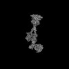 40917MC 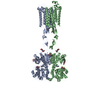 8szfC 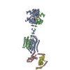 8szgC 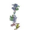 8szhC M: map data used to model this data C: citing same article ( |
|---|---|
| Similar structure data |


-Links
 PDBj
PDBj


- Assembly
| Deposited unit | 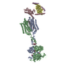
|
|---|---|
| 1 |
|
-Components
-Guanine nucleotide-binding protein ... , 3 types, 3 molecules CDE
| #1: Protein | Mass: 40584.156 Da / Num. of mol.: 1 / Mutation: S47N, G203A, E245A, A326S Source method: isolated from a genetically manipulated source Source: (gene. exp.) Homo sapiens (human) / Gene: GNAI3 / Production host:   Spodoptera frugiperda (fall armyworm) / References: UniProt: P08754 Spodoptera frugiperda (fall armyworm) / References: UniProt: P08754 |
|---|---|
| #2: Protein | Mass: 37573.988 Da / Num. of mol.: 1 Source method: isolated from a genetically manipulated source Source: (gene. exp.) Homo sapiens (human) / Gene: GNB1 / Production host: Spodoptera frugiperda (fall armyworm) / References: UniProt: P62873 |
| #3: Protein | Mass: 7861.143 Da / Num. of mol.: 1 Source method: isolated from a genetically manipulated source Source: (gene. exp.) Homo sapiens (human) / Gene: GNG2 / Production host: Spodoptera frugiperda (fall armyworm) / References: UniProt: P59768 |
-Extracellular calcium-sensing ... , 2 types, 2 molecules AB
| #4: Protein | Mass: 105938.789 Da / Num. of mol.: 1 Source method: isolated from a genetically manipulated source Source: (gene. exp.) Homo sapiens (human) / Gene: CASR, GPRC2A, PCAR1 / Production host: Spodoptera frugiperda (fall armyworm) / References: UniProt: P41180 |
|---|---|
| #5: Protein | Mass: 108023.055 Da / Num. of mol.: 1 Source method: isolated from a genetically manipulated source Source: (gene. exp.) Homo sapiens (human) / Gene: CASR, GPRC2A, PCAR1 / Production host: Spodoptera frugiperda (fall armyworm) / References: UniProt: P41180 |
-Sugars , 2 types, 6 molecules 
| #6: Polysaccharide | Source method: isolated from a genetically manipulated source #8: Sugar |  Type: D-saccharide, beta linking / Mass: 221.208 Da / Num. of mol.: 3 / Source method: obtained synthetically / Formula: C8H15NO6 Type: D-saccharide, beta linking / Mass: 221.208 Da / Num. of mol.: 3 / Source method: obtained synthetically / Formula: C8H15NO6 |
|---|
-Non-polymers , 5 types, 12 molecules 


| #7: Chemical |  Mass: 787.121 Da / Num. of mol.: 2 / Source method: obtained synthetically / Formula: C44H85NO8P / Feature type: SUBJECT OF INVESTIGATION / Comment: DOPC, phospholipid*YM Mass: 787.121 Da / Num. of mol.: 2 / Source method: obtained synthetically / Formula: C44H85NO8P / Feature type: SUBJECT OF INVESTIGATION / Comment: DOPC, phospholipid*YM#9: Chemical | ChemComp-CA /  Mass: 40.078 Da / Num. of mol.: 4 / Source method: obtained synthetically / Formula: Ca / Feature type: SUBJECT OF INVESTIGATION Mass: 40.078 Da / Num. of mol.: 4 / Source method: obtained synthetically / Formula: Ca / Feature type: SUBJECT OF INVESTIGATION#10: Chemical |  Type: L-peptide linking / Mass: 204.225 Da / Num. of mol.: 3 / Source method: obtained synthetically / Formula: C11H12N2O2 / Feature type: SUBJECT OF INVESTIGATION Type: L-peptide linking / Mass: 204.225 Da / Num. of mol.: 3 / Source method: obtained synthetically / Formula: C11H12N2O2 / Feature type: SUBJECT OF INVESTIGATION#11: Chemical |  Mass: 94.971 Da / Num. of mol.: 2 / Source method: obtained synthetically / Formula: PO4 / Feature type: SUBJECT OF INVESTIGATION Mass: 94.971 Da / Num. of mol.: 2 / Source method: obtained synthetically / Formula: PO4 / Feature type: SUBJECT OF INVESTIGATION#12: Chemical | ChemComp-CLR / |  Mass: 386.654 Da / Num. of mol.: 1 / Source method: obtained synthetically / Formula: C27H46O / Feature type: SUBJECT OF INVESTIGATION Mass: 386.654 Da / Num. of mol.: 1 / Source method: obtained synthetically / Formula: C27H46O / Feature type: SUBJECT OF INVESTIGATION |
|---|
-Details
| Has ligand of interest | Y |
|---|---|
| Has protein modification | Y |
-Experimental details
-Experiment
| Experiment | Method: ELECTRON MICROSCOPY |
|---|---|
| EM experiment | Aggregation state: PARTICLE / 3D reconstruction method: single particle reconstruction |
- Sample preparation
| Component | Name: PAM-free human calcium-sensing receptor CaSR-Gi complex in lipid nanodiscs Type: COMPLEX / Entity ID: #1-#5 / Source: RECOMBINANT |
|---|---|
| Source (natural) | Organism: Homo sapiens (human) |
| Source (recombinant) | Organism: Spodoptera frugiperda (fall armyworm) |
| Buffer solution | pH: 7.5 |
| Specimen | Embedding applied: NO / Shadowing applied: NO / Staining applied: NO / Vitrification applied: YES |
| Vitrification | Cryogen name: ETHANE |
- Electron microscopy imaging
| Experimental equipment |  Model: Titan Krios / Image courtesy: FEI Company |
|---|---|
| Microscopy | Model: FEI TITAN KRIOS |
| Electron gun | Electron source:  FIELD EMISSION GUN / Accelerating voltage: 300 kV / Illumination mode: FLOOD BEAM FIELD EMISSION GUN / Accelerating voltage: 300 kV / Illumination mode: FLOOD BEAM |
| Electron lens | Mode: BRIGHT FIELD / Nominal defocus max: 1500 nm / Nominal defocus min: 500 nm |
| Image recording | Electron dose: 50 e/Å2 / Film or detector model: GATAN K3 BIOQUANTUM (6k x 4k) |
- Processing
| EM software | Name: PHENIX / Version: 1.20.1_4487: / Category: model refinement | ||||||||||||||||||||||||
|---|---|---|---|---|---|---|---|---|---|---|---|---|---|---|---|---|---|---|---|---|---|---|---|---|---|
| CTF correction | Type: PHASE FLIPPING AND AMPLITUDE CORRECTION | ||||||||||||||||||||||||
| 3D reconstruction | Resolution: 3.5 Å / Resolution method: FSC 0.143 CUT-OFF / Num. of particles: 167678 / Symmetry type: POINT | ||||||||||||||||||||||||
| Refine LS restraints |
|
