 Movie
Movie Controller


+ Open data

- Basic information
| Entry | Database: PDB / ID: 8syg | ||||||
|---|---|---|---|---|---|---|---|
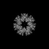
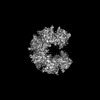
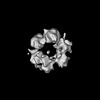
| Title | Cryo-EM structure of tetradecameric hub domain of CaMKII alpha | ||||||
 Components Components | Venus-tagged CaMKII Alpha Association Domain | ||||||
 Keywords Keywords | SIGNALING PROTEIN / High-order oligomer / Protein Kinase / Signaling / Memory | ||||||
| Function / homology |  Function and homology information Function and homology information: / HSF1-dependent transactivation / RAF activation / Ion transport by P-type ATPases / peptidyl-threonine autophosphorylation / calcium- and calmodulin-dependent protein kinase complex / regulation of endocannabinoid signaling pathway / neurotransmitter receptor transport to plasma membrane / Interferon gamma signaling / Ca2+/calmodulin-dependent protein kinase ...: / HSF1-dependent transactivation / RAF activation / Ion transport by P-type ATPases / peptidyl-threonine autophosphorylation / calcium- and calmodulin-dependent protein kinase complex / regulation of endocannabinoid signaling pathway / neurotransmitter receptor transport to plasma membrane / Interferon gamma signaling / Ca2+/calmodulin-dependent protein kinase / negative regulation of hydrolase activity / dendritic spine development / regulation of neurotransmitter secretion / Trafficking of AMPA receptors / positive regulation of calcium ion transport / regulation of neuron migration / calcium/calmodulin-dependent protein kinase activity / Ca2+ pathway / regulation of mitochondrial membrane permeability involved in apoptotic process / RAF/MAP kinase cascade / GTPase activating protein binding / Ion homeostasis / dendrite morphogenesis / regulation of neurotransmitter receptor localization to postsynaptic specialization membrane / negative regulation of ferroptosis / Unblocking of NMDA receptors, glutamate binding and activation / regulation of neuronal synaptic plasticity / glutamate receptor binding / postsynaptic cytosol / cell surface receptor signaling pathway via JAK-STAT / regulation of protein localization to plasma membrane / presynaptic cytosol / cellular response to interferon-beta / positive regulation of cardiac muscle cell apoptotic process / ionotropic glutamate receptor signaling pathway / dendrite cytoplasm / response to ischemia / bioluminescence / angiotensin-activated signaling pathway / positive regulation of receptor signaling pathway via JAK-STAT / generation of precursor metabolites and energy / G1/S transition of mitotic cell cycle / cellular response to type II interferon / Schaffer collateral - CA1 synapse / kinase activity / calcium ion transport / dendritic spine / calmodulin binding / neuron projection / postsynaptic density / protein serine kinase activity / axon / protein serine/threonine kinase activity / neuronal cell body / synapse / dendrite / glutamatergic synapse / protein homodimerization activity / mitochondrion / ATP binding / metal ion binding / identical protein binding / cytoplasm / cytosol Similarity search - Function | ||||||
| Biological species |   Aequorea victoria (jellyfish) Aequorea victoria (jellyfish) | ||||||
| Method | ELECTRON MICROSCOPY / single particle reconstruction / cryo EM / Resolution: 2.6 Å | ||||||
 Authors Authors | Chien, C.-T. / Chiu, W. / Khan, S. | ||||||
| Funding support |  United States, 1items United States, 1items
| ||||||
 Citation Citation | Journal: Commun Biol / Year: 2024 Title: Hub stability in the calcium calmodulin-dependent protein kinase II. Authors: Chih-Ta Chien / Henry Puhl / Steven S Vogel / Justin E Molloy / Wah Chiu / Shahid Khan /  Abstract: The calcium calmodulin protein kinase II (CaMKII) is a multi-subunit ring assembly with a central hub formed by the association domains. There is evidence for hub polymorphism between and within ...The calcium calmodulin protein kinase II (CaMKII) is a multi-subunit ring assembly with a central hub formed by the association domains. There is evidence for hub polymorphism between and within CaMKII isoforms, but the link between polymorphism and subunit exchange has not been resolved. Here, we present near-atomic resolution cryogenic electron microscopy (cryo-EM) structures revealing that hubs from the α and β isoforms, either standalone or within an β holoenzyme, coexist as 12 and 14 subunit assemblies. Single-molecule fluorescence microscopy of Venus-tagged holoenzymes detects intermediate assemblies and progressive dimer loss due to intrinsic holoenzyme lability, and holoenzyme disassembly into dimers upon mutagenesis of a conserved inter-domain contact. Molecular dynamics (MD) simulations show the flexibility of 4-subunit precursors, extracted in-silico from the β hub polymorphs, encompassing the curvature of both polymorphs. The MD explains how an open hub structure also obtained from the β holoenzyme sample could be created by dimer loss and analysis of its cryo-EM dataset reveals how the gap could open further. An assembly model, considering dimer concentration dependence and strain differences between polymorphs, proposes a mechanism for intrinsic hub lability to fine-tune the stoichiometry of αβ heterooligomers for their dynamic localization within synapses in neurons. | ||||||
| History |
|
- Structure visualization
| Structure viewer | Molecule: MolmilJmol/JSmol |
|---|
- Downloads & links
-Download
| PDBx/mmCIF format | 8syg.cif.gz | 711.8 KB | Display | PDBx/mmCIF format |
|---|---|---|---|---|
| PDB format | pdb8syg.ent.gz | 585.5 KB | Display | PDB format |
| PDBx/mmJSON format | 8syg.json.gz | Tree view | PDBx/mmJSON format | |
| Others |  Other downloads Other downloads |
-Validation report
| Arichive directory | https://data.pdbj.org/pub/pdb/validation_reports/sy/8sygftp://data.pdbj.org/pub/pdb/validation_reports/sy/8syg | HTTPS FTP |
|---|
-Related structure data
| Related structure data | 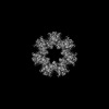 40873MC 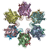 8t15C  8t17C  8t18C 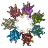 8t6kC 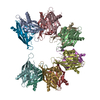 8t6qC M: map data used to model this data C: citing same article ( |
|---|---|
| Similar structure data |


-Links
 PDBj
PDBj


- Assembly
| Deposited unit | 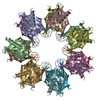
|
|---|---|
| 1 |
|
-Components
| #1: Protein | Mass: 45877.539 Da / Num. of mol.: 14 Source method: isolated from a genetically manipulated source Source: (gene. exp.) Aequorea victoria (jellyfish), (gene. exp.) Gene: Camk2a / Production host:  References: UniProt: P42212, UniProt: P11275, Ca2+/calmodulin-dependent protein kinase |
|---|
-Experimental details
-Experiment
| Experiment | Method: ELECTRON MICROSCOPY |
|---|---|
| EM experiment | Aggregation state: PARTICLE / 3D reconstruction method: single particle reconstruction |
- Sample preparation
| Component | Name: Venus-tagged CaMKII Alpha Association Domain / Type: COMPLEX / Entity ID: all / Source: RECOMBINANT |
|---|---|
| Molecular weight | Experimental value: NO |
| Source (natural) | Organism: |
| Source (recombinant) | Organism: |
| Buffer solution | pH: 7.5 |
| Specimen | Conc.: 20 mg/ml / Embedding applied: NO / Shadowing applied: NO / Staining applied: NO / Vitrification applied: YES |
| Vitrification | Cryogen name: ETHANE / Humidity: 100 % / Chamber temperature: 277 K |
- Electron microscopy imaging
| Experimental equipment |  Model: Titan Krios / Image courtesy: FEI Company |
|---|---|
| Microscopy | Model: FEI TITAN KRIOS |
| Electron gun | Electron source:  FIELD EMISSION GUN / Accelerating voltage: 300 kV / Illumination mode: FLOOD BEAM FIELD EMISSION GUN / Accelerating voltage: 300 kV / Illumination mode: FLOOD BEAM |
| Electron lens | Mode: BRIGHT FIELD / Nominal defocus max: 1500 nm / Nominal defocus min: 600 nm |
| Image recording | Electron dose: 60 e/Å2 / Film or detector model: FEI FALCON IV (4k x 4k) |
- Processing
| CTF correction | Type: PHASE FLIPPING AND AMPLITUDE CORRECTION |
|---|---|
| 3D reconstruction | Resolution: 2.6 Å / Resolution method: FSC 0.143 CUT-OFF / Num. of particles: 205902 / Symmetry type: POINT |
