 Movie
Movie Controller
 Structure viewers
 About Yorodumi Papers


+Search query
-Structure paper
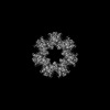
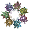
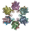
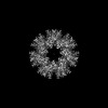
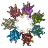
| Title | Hub stability in the calcium calmodulin-dependent protein kinase II. |
|---|---|
| Journal, issue, pages | Commun Biol, Vol. 7, Issue 1, Page 766, Year 2024 |
| Publish date | Jun 25, 2024 |
 Authors Authors | Chih-Ta Chien / Henry Puhl / Steven S Vogel / Justin E Molloy / Wah Chiu / Shahid Khan /   |
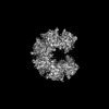
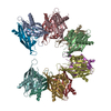
| PubMed Abstract | The calcium calmodulin protein kinase II (CaMKII) is a multi-subunit ring assembly with a central hub formed by the association domains. There is evidence for hub polymorphism between and within ...The calcium calmodulin protein kinase II (CaMKII) is a multi-subunit ring assembly with a central hub formed by the association domains. There is evidence for hub polymorphism between and within CaMKII isoforms, but the link between polymorphism and subunit exchange has not been resolved. Here, we present near-atomic resolution cryogenic electron microscopy (cryo-EM) structures revealing that hubs from the α and β isoforms, either standalone or within an β holoenzyme, coexist as 12 and 14 subunit assemblies. Single-molecule fluorescence microscopy of Venus-tagged holoenzymes detects intermediate assemblies and progressive dimer loss due to intrinsic holoenzyme lability, and holoenzyme disassembly into dimers upon mutagenesis of a conserved inter-domain contact. Molecular dynamics (MD) simulations show the flexibility of 4-subunit precursors, extracted in-silico from the β hub polymorphs, encompassing the curvature of both polymorphs. The MD explains how an open hub structure also obtained from the β holoenzyme sample could be created by dimer loss and analysis of its cryo-EM dataset reveals how the gap could open further. An assembly model, considering dimer concentration dependence and strain differences between polymorphs, proposes a mechanism for intrinsic hub lability to fine-tune the stoichiometry of αβ heterooligomers for their dynamic localization within synapses in neurons. |
 External links External links | Commun Biol / PubMed:38918547 / PubMed Central |
| Methods | EM (single particle) |
| Resolution | 2.6 - 8.4 Å |
| Structure data | EMDB-40873, PDB-8syg: EMDB-40955, PDB-8t15: EMDB-40956, PDB-8t17: EMDB-40957, PDB-8t18: EMDB-41070, PDB-8t6k: EMDB-41077, PDB-8t6q: 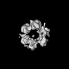 EMDB-43385: Cryo-EM map of close dodecameric CaMKII beta holoenzyme T287A T306A T307A |
| Source |
|
 Keywords Keywords | SIGNALING PROTEIN / High-order oligomer / Protein Kinase / Signaling / Memory |


 aequorea victoria (jellyfish)