+ Open data
- Basic information
| Entry | Database: PDB / ID: 8sur | |||||||||
|---|---|---|---|---|---|---|---|---|---|---|
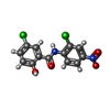
| Title | TMEM16F bound with Niclosamide | |||||||||
 Components Components | Anoctamin-6 | |||||||||
 Keywords Keywords | MEMBRANE PROTEIN / Ca2+-activated ion channels and lipid scramblases | |||||||||
| Function / homology |  Function and homology information Function and homology informationcalcium activated phospholipid scrambling / calcium activated galactosylceramide scrambling / calcium activated phosphatidylserine scrambling / calcium activated phosphatidylcholine scrambling / positive regulation of potassium ion export across plasma membrane / positive regulation of monoatomic ion transmembrane transport / purinergic nucleotide receptor signaling pathway / phospholipid scramblase activity / cholinergic synapse / bone mineralization involved in bone maturation ...calcium activated phospholipid scrambling / calcium activated galactosylceramide scrambling / calcium activated phosphatidylserine scrambling / calcium activated phosphatidylcholine scrambling / positive regulation of potassium ion export across plasma membrane / positive regulation of monoatomic ion transmembrane transport / purinergic nucleotide receptor signaling pathway / phospholipid scramblase activity / cholinergic synapse / bone mineralization involved in bone maturation / negative regulation of cell volume / intracellularly calcium-gated chloride channel activity / plasma membrane phospholipid scrambling / voltage-gated monoatomic ion channel activity / positive regulation of phagocytosis, engulfment / bleb assembly / Stimuli-sensing channels / calcium-activated cation channel activity / positive regulation of monocyte chemotaxis / chloride transport / dendritic cell chemotaxis / phospholipid translocation / regulation of postsynaptic membrane potential / positive regulation of bone mineralization / chloride channel complex / Neutrophil degranulation / chloride transmembrane transport / synaptic membrane / establishment of localization in cell / calcium ion transmembrane transport / blood coagulation / positive regulation of apoptotic process / protein homodimerization activity / metal ion binding / identical protein binding / plasma membrane Similarity search - Function | |||||||||
| Biological species |  | |||||||||
| Method | ELECTRON MICROSCOPY / single particle reconstruction / Resolution: 3.1 Å | |||||||||
 Authors Authors | Feng, S. / Cheng, Y. | |||||||||
| Funding support |  United States, 1items United States, 1items
| |||||||||
 Citation Citation |  Journal: Nat Commun / Year: 2023 Journal: Nat Commun / Year: 2023Title: Identification of a drug binding pocket in TMEM16F calcium-activated ion channel and lipid scramblase. Authors: Shengjie Feng / Cristina Puchades / Juyeon Ko / Hao Wu / Yifei Chen / Eric E Figueroa / Shuo Gu / Tina W Han / Brandon Ho / Tong Cheng / Junrui Li / Brian Shoichet / Yuh Nung Jan / Yifan Cheng / Lily Yeh Jan /  Abstract: The dual functions of TMEM16F as Ca-activated ion channel and lipid scramblase raise intriguing questions regarding their molecular basis. Intrigued by the ability of the FDA-approved drug ...The dual functions of TMEM16F as Ca-activated ion channel and lipid scramblase raise intriguing questions regarding their molecular basis. Intrigued by the ability of the FDA-approved drug niclosamide to inhibit TMEM16F-dependent syncytia formation induced by SARS-CoV-2, we examined cryo-EM structures of TMEM16F with or without bound niclosamide or 1PBC, a known blocker of TMEM16A Ca-activated Cl channel. Here, we report evidence for a lipid scrambling pathway along a groove harboring a lipid trail outside the ion permeation pore. This groove contains the binding pocket for niclosamide and 1PBC. Mutations of two residues in this groove specifically affect lipid scrambling. Whereas mutations of some residues in the binding pocket of niclosamide and 1PBC reduce their inhibition of TMEM16F-mediated Ca influx and PS exposure, other mutations preferentially affect the ability of niclosamide and/or 1PBC to inhibit TMEM16F-mediated PS exposure, providing further support for separate pathways for ion permeation and lipid scrambling. | |||||||||
| History |
|
- Structure visualization
| Structure viewer | Molecule:  Molmil Molmil Jmol/JSmol Jmol/JSmol |
|---|
- Downloads & links
- Download
| PDBx/mmCIF format |  8sur.cif.gz 8sur.cif.gz | 477.6 KB | Display |  PDBx/mmCIF format PDBx/mmCIF format |
|---|---|---|---|---|
| PDB format |  pdb8sur.ent.gz pdb8sur.ent.gz | 391.4 KB | Display |  PDB format PDB format |
| PDBx/mmJSON format |  8sur.json.gz 8sur.json.gz | Tree view |  PDBx/mmJSON format PDBx/mmJSON format | |
| Others |  Other downloads Other downloads |
-Validation report
| Arichive directory |  https://data.pdbj.org/pub/pdb/validation_reports/su/8sur https://data.pdbj.org/pub/pdb/validation_reports/su/8sur ftp://data.pdbj.org/pub/pdb/validation_reports/su/8sur ftp://data.pdbj.org/pub/pdb/validation_reports/su/8sur | HTTPS FTP |
|---|
-Related structure data
| Related structure data |  40776MC  8sunC  8tagC  8taiC  8talC M: map data used to model this data C: citing same article ( |
|---|---|
| Similar structure data | Similarity search - Function & homology  F&H Search F&H Search |
- Links
Links
- Assembly
| Deposited unit | 
|
|---|---|
| 1 | 
|
- Components
| #1: Protein | Mass: 106367.727 Da / Num. of mol.: 2 Source method: isolated from a genetically manipulated source Source: (gene. exp.)   Homo sapiens (human) / References: UniProt: Q6P9J9 Homo sapiens (human) / References: UniProt: Q6P9J9#2: Polysaccharide | #3: Chemical | ChemComp-CA / #4: Sugar | #5: Chemical | ChemComp-VUT / | Has ligand of interest | Y | Has protein modification | Y | |
|---|
-Experimental details
-Experiment
| Experiment | Method: ELECTRON MICROSCOPY |
|---|---|
| EM experiment | Aggregation state: CELL / 3D reconstruction method: single particle reconstruction |
- Sample preparation
| Component | Name: 16F / Type: COMPLEX / Entity ID: #1 / Source: RECOMBINANT |
|---|---|
| Source (natural) | Organism:  |
| Source (recombinant) | Organism:  Homo sapiens (human) Homo sapiens (human) |
| Buffer solution | pH: 7.5 |
| Specimen | Embedding applied: NO / Shadowing applied: NO / Staining applied: NO / Vitrification applied: NO |
- Electron microscopy imaging
| Experimental equipment |  Model: Titan Krios / Image courtesy: FEI Company |
|---|---|
| Microscopy | Model: FEI TITAN KRIOS |
| Electron gun | Electron source:  FIELD EMISSION GUN / Accelerating voltage: 300 kV / Illumination mode: SPOT SCAN FIELD EMISSION GUN / Accelerating voltage: 300 kV / Illumination mode: SPOT SCAN |
| Electron lens | Mode: BRIGHT FIELD / Nominal defocus max: 2000 nm / Nominal defocus min: 1000 nm |
| Image recording | Electron dose: 66 e/Å2 / Film or detector model: GATAN K3 (6k x 4k) |
- Processing
| CTF correction | Type: NONE |
|---|---|
| 3D reconstruction | Resolution: 3.1 Å / Resolution method: FSC 0.143 CUT-OFF / Num. of particles: 1047816 / Symmetry type: POINT |
 Movie
Movie Controller


 PDBj
PDBj


