 Movie
Movie Controller

[English] 日本語
 Yorodumi
- PDB-8sm0: Crystal structure of human complement receptor 2 (CD21) in comple... -
+ Open data

- Basic information
| Entry | Database: PDB / ID: 8sm0 | ||||||
|---|---|---|---|---|---|---|---|
| Title | Crystal structure of human complement receptor 2 (CD21) in complex with Epstein-Barr virus major glycoprotein gp350 | ||||||
 Components Components |
| ||||||
 Keywords Keywords | VIRAL PROTEIN/IMMUNE SYSTEM / VIRAL PROTEIN / VIRAL PROTEIN-IMMUNE SYSTEM complex | ||||||
| Function / homology |  Function and homology information Function and homology informationnegative regulation of complement activation, classical pathway / complement receptor activity / complement binding / T cell mediated immunity / complement activation, alternative pathway / immunoglobulin receptor binding / type I interferon-mediated signaling pathway / B cell activation / B cell proliferation / host cell membrane ...negative regulation of complement activation, classical pathway / complement receptor activity / complement binding / T cell mediated immunity / complement activation, alternative pathway / immunoglobulin receptor binding / type I interferon-mediated signaling pathway / B cell activation / B cell proliferation / host cell membrane / complement activation, classical pathway / B cell differentiation / Regulation of Complement cascade / transmembrane signaling receptor activity / virus receptor activity / receptor complex / immune response / receptor ligand activity / symbiont entry into host cell / virion membrane / protein homodimerization activity / extracellular space / DNA binding / extracellular exosome / membrane / plasma membrane Similarity search - Function | ||||||
| Biological species |  Homo sapiens (human) Homo sapiens (human) Human herpesvirus 4 (Epstein-Barr virus) Human herpesvirus 4 (Epstein-Barr virus) | ||||||
| Method |  X-RAY DIFFRACTION / SYNCHROTRON / MOLECULAR REPLACEMENT / Resolution: 1.68 Å X-RAY DIFFRACTION / SYNCHROTRON / MOLECULAR REPLACEMENT / Resolution: 1.68 Å | ||||||
 Authors Authors | Chen, W.-H. / Bu, W. / Cohen, J.I. / Kanekiyo, M. / Joyce, M.G. | ||||||
| Funding support |  United States, 1items United States, 1items
| ||||||
 Citation Citation | Journal: Immunity / Year: 2025 Title: Structural basis for complement receptor engagement and virus neutralization through Epstein-Barr virus gp350. Authors: Joyce, M.G. / Bu, W. / Chen, W.H. / Gillespie, R.A. / Andrews, S.F. / Wheatley, A.K. / Tsybovsky, Y. / Jensen, J.L. / Stephens, T. / Prabhakaran, M. / Fisher, B.E. / Narpala, S.R. / Bagchi, ...Authors: Joyce, M.G. / Bu, W. / Chen, W.H. / Gillespie, R.A. / Andrews, S.F. / Wheatley, A.K. / Tsybovsky, Y. / Jensen, J.L. / Stephens, T. / Prabhakaran, M. / Fisher, B.E. / Narpala, S.R. / Bagchi, M. / McDermott, A.B. / Nabel, G.J. / Kwong, P.D. / Mascola, J.R. / Cohen, J.I. / Kanekiyo, M. | ||||||
| History |
|
- Structure visualization
| Structure viewer | Molecule: MolmilJmol/JSmol |
|---|
- Downloads & links
-Download
| PDBx/mmCIF format | 8sm0.cif.gz | 320 KB | Display | PDBx/mmCIF format |
|---|---|---|---|---|
| PDB format | pdb8sm0.ent.gz | 265.4 KB | Display | PDB format |
| PDBx/mmJSON format | 8sm0.json.gz | Tree view | PDBx/mmJSON format | |
| Others |  Other downloads Other downloads |
-Validation report
| Arichive directory | https://data.pdbj.org/pub/pdb/validation_reports/sm/8sm0ftp://data.pdbj.org/pub/pdb/validation_reports/sm/8sm0 | HTTPS FTP |
|---|
-Related structure data
| Related structure data |  8sefC  8sgaC  8sggC  8sgnC  8sicC 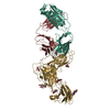 8sm1C  1ly2S  2h6oS S: Starting model for refinement C: citing same article ( |
|---|---|
| Similar structure data |
-Links
 PDBj
PDBj

- Assembly
| Deposited unit | 
| ||||||||
|---|---|---|---|---|---|---|---|---|---|
| 1 |
| ||||||||
| Unit cell |
|
-Components
| #1: Protein | Mass: 14155.256 Da / Num. of mol.: 1 / Fragment: first two amino-terminal domains Source method: isolated from a genetically manipulated source Source: (gene. exp.) Homo sapiens (human) / Gene: CR2, C3DR / Production host: Homo sapiens (human) / References: UniProt: P20023 | ||||||||
|---|---|---|---|---|---|---|---|---|---|
| #2: Protein | Mass: 46895.230 Da / Num. of mol.: 1 Source method: isolated from a genetically manipulated source Source: (gene. exp.) Human herpesvirus 4 (Epstein-Barr virus)Strain: B95-8 / Gene: BLLF1 / Production host: Homo sapiens (human) / References: UniProt: P03200 | ||||||||
| #3: Sugar | ChemComp-NAG /   Type: D-saccharide, beta linking / Mass: 221.208 Da / Num. of mol.: 12 Type: D-saccharide, beta linking / Mass: 221.208 Da / Num. of mol.: 12Source method: isolated from a genetically manipulated source Formula: C8H15NO6 #4: Chemical | ChemComp-SO4 / |   Mass: 96.063 Da / Num. of mol.: 1 / Source method: obtained synthetically / Formula: SO4 Mass: 96.063 Da / Num. of mol.: 1 / Source method: obtained synthetically / Formula: SO4#5: Water | ChemComp-HOH / |  Mass: 18.015 Da / Num. of mol.: 237 / Source method: isolated from a natural source / Formula: H2O Mass: 18.015 Da / Num. of mol.: 237 / Source method: isolated from a natural source / Formula: H2OHas ligand of interest | N | Has protein modification | Y | |
-Experimental details
-Experiment
| Experiment | Method: X-RAY DIFFRACTION / Number of used crystals: 1 |
|---|
- Sample preparation
| Crystal | Density Matthews: 3.03 Å3/Da / Density % sol: 59.44 % |
|---|---|
| Crystal grow | Temperature: 291 K / Method: vapor diffusion, hanging drop Details: 1.60M AMMONIUM SULFATE, 2% PEG 400, 0.5% GLYCEROL, 0.1M SODIUM ACETATE PH5.5 |
-Data collection
| Diffraction | Mean temperature: 100 K / Serial crystal experiment: N |
|---|---|
| Diffraction source | Source: SYNCHROTRON / Site: APS / Beamline: 24-ID-E / Wavelength: 0.98 Å |
| Detector | Type: DECTRIS EIGER X 16M / Detector: PIXEL / Date: Nov 18, 2017 |
| Radiation | Protocol: SINGLE WAVELENGTH / Monochromatic (M) / Laue (L): M / Scattering type: x-ray |
| Radiation wavelength | Wavelength: 0.98 Å / Relative weight: 1 |
| Reflection | Resolution: 1.68→50 Å / Num. obs: 56365 / % possible obs: 84.7 % / Redundancy: 3.1 % / Rmerge(I) obs: 0.08 / Net I/σ(I): 10.3 |
| Reflection shell | Resolution: 1.684→1.75 Å / Rmerge(I) obs: 0.744 / Mean I/σ(I) obs: 1.8 / Num. unique obs: 1994 |
- Processing
| Software |
| ||||||||||||||||||||||||||||||||||||||||||||||||||||||||||||||||||||||||||||||||||||||||||||||||||||||||||||||||||||||||||||||||||||||||||||||||||||||||||||||||||||||||||||||||||||||||||||||||||||||||||||||||||||||||||||||||||||||||||||||||||||||||||
|---|---|---|---|---|---|---|---|---|---|---|---|---|---|---|---|---|---|---|---|---|---|---|---|---|---|---|---|---|---|---|---|---|---|---|---|---|---|---|---|---|---|---|---|---|---|---|---|---|---|---|---|---|---|---|---|---|---|---|---|---|---|---|---|---|---|---|---|---|---|---|---|---|---|---|---|---|---|---|---|---|---|---|---|---|---|---|---|---|---|---|---|---|---|---|---|---|---|---|---|---|---|---|---|---|---|---|---|---|---|---|---|---|---|---|---|---|---|---|---|---|---|---|---|---|---|---|---|---|---|---|---|---|---|---|---|---|---|---|---|---|---|---|---|---|---|---|---|---|---|---|---|---|---|---|---|---|---|---|---|---|---|---|---|---|---|---|---|---|---|---|---|---|---|---|---|---|---|---|---|---|---|---|---|---|---|---|---|---|---|---|---|---|---|---|---|---|---|---|---|---|---|---|---|---|---|---|---|---|---|---|---|---|---|---|---|---|---|---|---|---|---|---|---|---|---|---|---|---|---|---|---|---|---|---|---|---|---|---|---|---|---|---|---|---|---|---|---|---|---|---|---|
| Refinement | Method to determine structure: MOLECULAR REPLACEMENT Starting model: 2H6O, 1LY2 Resolution: 1.68→30.68 Å / SU ML: 0.19 / Cross valid method: FREE R-VALUE / σ(F): 1.35 / Phase error: 32.54
| ||||||||||||||||||||||||||||||||||||||||||||||||||||||||||||||||||||||||||||||||||||||||||||||||||||||||||||||||||||||||||||||||||||||||||||||||||||||||||||||||||||||||||||||||||||||||||||||||||||||||||||||||||||||||||||||||||||||||||||||||||||||||||
| Solvent computation | Shrinkage radii: 0.9 Å / VDW probe radii: 1.11 Å | ||||||||||||||||||||||||||||||||||||||||||||||||||||||||||||||||||||||||||||||||||||||||||||||||||||||||||||||||||||||||||||||||||||||||||||||||||||||||||||||||||||||||||||||||||||||||||||||||||||||||||||||||||||||||||||||||||||||||||||||||||||||||||
| Displacement parameters | Biso mean: 61.73 Å2 | ||||||||||||||||||||||||||||||||||||||||||||||||||||||||||||||||||||||||||||||||||||||||||||||||||||||||||||||||||||||||||||||||||||||||||||||||||||||||||||||||||||||||||||||||||||||||||||||||||||||||||||||||||||||||||||||||||||||||||||||||||||||||||
| Refinement step | Cycle: LAST / Resolution: 1.68→30.68 Å
| ||||||||||||||||||||||||||||||||||||||||||||||||||||||||||||||||||||||||||||||||||||||||||||||||||||||||||||||||||||||||||||||||||||||||||||||||||||||||||||||||||||||||||||||||||||||||||||||||||||||||||||||||||||||||||||||||||||||||||||||||||||||||||
| LS refinement shell |
| ||||||||||||||||||||||||||||||||||||||||||||||||||||||||||||||||||||||||||||||||||||||||||||||||||||||||||||||||||||||||||||||||||||||||||||||||||||||||||||||||||||||||||||||||||||||||||||||||||||||||||||||||||||||||||||||||||||||||||||||||||||||||||
| Refinement TLS params. | Method: refined / Refine-ID: X-RAY DIFFRACTION
| ||||||||||||||||||||||||||||||||||||||||||||||||||||||||||||||||||||||||||||||||||||||||||||||||||||||||||||||||||||||||||||||||||||||||||||||||||||||||||||||||||||||||||||||||||||||||||||||||||||||||||||||||||||||||||||||||||||||||||||||||||||||||||
| Refinement TLS group |
|
