 Movie
Movie Controller

[English] 日本語
 Yorodumi
- PDB-8sip: Structure of a mouse IgG antibody fragment that binds Inosine, an... -
+ Open data

- Basic information
| Entry | Database: PDB / ID: 8sip | ||||||
|---|---|---|---|---|---|---|---|
| Title | Structure of a mouse IgG antibody fragment that binds Inosine, an RNA modification | ||||||
 Components Components |
| ||||||
 Keywords Keywords | IMMUNE SYSTEM / IgG Fab / Inosine / RNA binding protein / modified RNA / small molecule ligand | ||||||
| Function / homology | INOSINE Function and homology information Function and homology information | ||||||
| Biological species |  | ||||||
| Method |  X-RAY DIFFRACTION / SYNCHROTRON / MOLECULAR REPLACEMENT / Resolution: 1.94 Å X-RAY DIFFRACTION / SYNCHROTRON / MOLECULAR REPLACEMENT / Resolution: 1.94 Å | ||||||
 Authors Authors | Aoki, S.T. | ||||||
| Funding support | 1items
| ||||||
 Citation Citation | Journal: Nucleic Acids Res. / Year: 2025 Title: In silico lambda-dynamics predicts protein binding specificities to modified RNAs. Authors: Angelo, M. / Zhang, W. / Vilseck, J.Z. / Aoki, S.T. | ||||||
| History |
|
- Structure visualization
| Structure viewer | Molecule: MolmilJmol/JSmol |
|---|
- Downloads & links
-Download
| PDBx/mmCIF format | 8sip.cif.gz | 209.6 KB | Display | PDBx/mmCIF format |
|---|---|---|---|---|
| PDB format | pdb8sip.ent.gz | 145.2 KB | Display | PDB format |
| PDBx/mmJSON format | 8sip.json.gz | Tree view | PDBx/mmJSON format | |
| Others |  Other downloads Other downloads |
-Validation report
| Arichive directory | https://data.pdbj.org/pub/pdb/validation_reports/si/8sipftp://data.pdbj.org/pub/pdb/validation_reports/si/8sip | HTTPS FTP |
|---|
-Related structure data
| Related structure data | 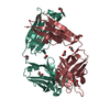 8tcaC 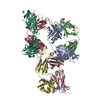 8vevC C: citing same article ( |
|---|---|
| Similar structure data | |
| Experimental dataset #1 | Data reference: 10.18430/M38SIP / Data set type: diffraction image data |
-Links
 PDBj
PDBj

- Assembly
| Deposited unit | 
| ||||||||||||
|---|---|---|---|---|---|---|---|---|---|---|---|---|---|
| 1 |
| ||||||||||||
| Unit cell |
|
-Components
| #1: Antibody | Mass: 23942.006 Da / Num. of mol.: 1 Source method: isolated from a genetically manipulated source Source: (gene. exp.)  Homo sapiens (human) Homo sapiens (human) |
|---|---|
| #2: Antibody | Mass: 24295.979 Da / Num. of mol.: 1 Source method: isolated from a genetically manipulated source Source: (gene. exp.) Homo sapiens (human) |
| #3: Chemical | ChemComp-NOS / 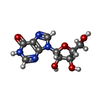  Mass: 268.226 Da / Num. of mol.: 1 / Source method: obtained synthetically / Formula: C10H12N4O5 / Feature type: SUBJECT OF INVESTIGATION Mass: 268.226 Da / Num. of mol.: 1 / Source method: obtained synthetically / Formula: C10H12N4O5 / Feature type: SUBJECT OF INVESTIGATION |
| #4: Water | ChemComp-HOH /  Mass: 18.015 Da / Num. of mol.: 235 / Source method: isolated from a natural source / Formula: H2O Mass: 18.015 Da / Num. of mol.: 235 / Source method: isolated from a natural source / Formula: H2O |
| Has ligand of interest | Y |
| Has protein modification | Y |
-Experimental details
-Experiment
| Experiment | Method: X-RAY DIFFRACTION / Number of used crystals: 1 |
|---|
- Sample preparation
| Crystal | Density Matthews: 2.31 Å3/Da / Density % sol: 46.82 % |
|---|---|
| Crystal grow | Temperature: 293 K / Method: vapor diffusion, sitting drop / pH: 8.3 / Details: 50 mM TRIS PH 8.3, 15% PEG 4000, 0.1 mM EDTA |
-Data collection
| Diffraction | Mean temperature: 100 K / Serial crystal experiment: N |
|---|---|
| Diffraction source | Source: SYNCHROTRON / Site: APS  / Beamline: 31-ID / Wavelength: 0.9793 Å / Beamline: 31-ID / Wavelength: 0.9793 Å |
| Detector | Type: DECTRIS PILATUS3 S 6M / Detector: PIXEL / Date: Mar 31, 2023 |
| Radiation | Monochromator: Kohzu HLD-15 Double Crystal / Protocol: SINGLE WAVELENGTH / Monochromatic (M) / Laue (L): M / Scattering type: x-ray |
| Radiation wavelength | Wavelength: 0.9793 Å / Relative weight: 1 |
| Reflection | Resolution: 1.94→57.04 Å / Num. obs: 30888 / % possible obs: 96.2 % / Redundancy: 7.1 % / Biso Wilson estimate: 26.61 Å2 / CC1/2: 0.991 / CC star: 0.998 / Rmerge(I) obs: 0.1901 / Rpim(I) all: 0.06938 / Rrim(I) all: 0.2027 / Net I/σ(I): 6.33 |
| Reflection shell | Resolution: 1.94→2.009 Å / Redundancy: 4.9 % / Rmerge(I) obs: 1.506 / Mean I/σ(I) obs: 3.17 / Num. unique obs: 2931 / CC1/2: 0.586 / CC star: 0.86 / Rpim(I) all: 0.6427 / Rrim(I) all: 1.642 / % possible all: 91.25 |
- Processing
| Software |
| ||||||||||||||||||||||||||||||||||||||||||||||||||||||||||||||||||||||||||||||||||||
|---|---|---|---|---|---|---|---|---|---|---|---|---|---|---|---|---|---|---|---|---|---|---|---|---|---|---|---|---|---|---|---|---|---|---|---|---|---|---|---|---|---|---|---|---|---|---|---|---|---|---|---|---|---|---|---|---|---|---|---|---|---|---|---|---|---|---|---|---|---|---|---|---|---|---|---|---|---|---|---|---|---|---|---|---|---|
| Refinement | Method to determine structure: MOLECULAR REPLACEMENT / Resolution: 1.94→57.04 Å / SU ML: 0.242 / Cross valid method: FREE R-VALUE / σ(F): 1.96 / Phase error: 26.878 Stereochemistry target values: GeoStd + Monomer Library + CDL v1.2
| ||||||||||||||||||||||||||||||||||||||||||||||||||||||||||||||||||||||||||||||||||||
| Solvent computation | Shrinkage radii: 0.9 Å / VDW probe radii: 1.1 Å / Solvent model: FLAT BULK SOLVENT MODEL | ||||||||||||||||||||||||||||||||||||||||||||||||||||||||||||||||||||||||||||||||||||
| Displacement parameters | Biso mean: 32.66 Å2 | ||||||||||||||||||||||||||||||||||||||||||||||||||||||||||||||||||||||||||||||||||||
| Refinement step | Cycle: LAST / Resolution: 1.94→57.04 Å
| ||||||||||||||||||||||||||||||||||||||||||||||||||||||||||||||||||||||||||||||||||||
| Refine LS restraints |
| ||||||||||||||||||||||||||||||||||||||||||||||||||||||||||||||||||||||||||||||||||||
| LS refinement shell |
| ||||||||||||||||||||||||||||||||||||||||||||||||||||||||||||||||||||||||||||||||||||
| Refinement TLS params. | Method: refined / Origin x: -12.0772151881 Å / Origin y: 23.4036181553 Å / Origin z: -3.77583644921 Å
| ||||||||||||||||||||||||||||||||||||||||||||||||||||||||||||||||||||||||||||||||||||
| Refinement TLS group | Selection details: all |
