Movie
Movie Controller


+ Open data

- Basic information
| Entry | Database: PDB / ID: 8r3g | |||||||||
|---|---|---|---|---|---|---|---|---|---|---|
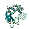
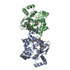
| Title | Central glycolytic genes regulator (CggR) bound to DNA operator | |||||||||
 Components Components |
| |||||||||
 Keywords Keywords | DNA BINDING PROTEIN / Bacillus subtilis / transcription repressor / glucose catabolism / SorC family | |||||||||
| Function / homology |  Function and homology information Function and homology informationregulation of DNA-templated transcription initiation / cis-regulatory region sequence-specific DNA binding / carbohydrate binding Similarity search - Function | |||||||||
| Biological species |  | |||||||||
| Method | ELECTRON MICROSCOPY / single particle reconstruction / cryo EM / Resolution: 4.4 Å | |||||||||
 Authors Authors | Skerlova, J. / Soltysova, M. / Rezacova, P. / Skubnik, K. | |||||||||
| Funding support |  Czech Republic, European Union, 2items Czech Republic, European Union, 2items
| |||||||||
 Citation Citation | Journal: Nucleic Acids Res / Year: 2024 Title: Structural characterization of two prototypical repressors of SorC family reveals tetrameric assemblies on DNA and mechanism of function. Authors: Markéta Šoltysová / Jana Škerlová / Petr Pachl / Karel Škubník / Milan Fábry / Irena Sieglová / Martina Farolfi / Irina Grishkovskaya / Michal Babiak / Jiří Nováček / Libor ...Authors: Markéta Šoltysová / Jana Škerlová / Petr Pachl / Karel Škubník / Milan Fábry / Irena Sieglová / Martina Farolfi / Irina Grishkovskaya / Michal Babiak / Jiří Nováček / Libor Krásný / Pavlína Řezáčová /  Abstract: The SorC family of transcriptional regulators plays a crucial role in controlling the carbohydrate metabolism and quorum sensing. We employed an integrative approach combining X-ray crystallography ...The SorC family of transcriptional regulators plays a crucial role in controlling the carbohydrate metabolism and quorum sensing. We employed an integrative approach combining X-ray crystallography and cryo-electron microscopy to investigate architecture and functional mechanism of two prototypical representatives of two sub-classes of the SorC family: DeoR and CggR from Bacillus subtilis. Despite possessing distinct DNA-binding domains, both proteins form similar tetrameric assemblies when bound to their respective DNA operators. Structural analysis elucidates the process by which the CggR-regulated gapA operon is derepressed through the action of two effectors: fructose-1,6-bisphosphate and newly confirmed dihydroxyacetone phosphate. Our findings provide the first comprehensive understanding of the DNA binding mechanism of the SorC-family proteins, shedding new light on their functional characteristics. | |||||||||
| History |
|
- Structure visualization
| Structure viewer | Molecule: MolmilJmol/JSmol |
|---|
- Downloads & links
-Download
| PDBx/mmCIF format | 8r3g.cif.gz | 302.8 KB | Display | PDBx/mmCIF format |
|---|---|---|---|---|
| PDB format | pdb8r3g.ent.gz | 238.9 KB | Display | PDB format |
| PDBx/mmJSON format | 8r3g.json.gz | Tree view | PDBx/mmJSON format | |
| Others |  Other downloads Other downloads |
-Validation report
| Arichive directory | https://data.pdbj.org/pub/pdb/validation_reports/r3/8r3gftp://data.pdbj.org/pub/pdb/validation_reports/r3/8r3g | HTTPS FTP |
|---|
-Related structure data
| Related structure data | 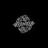 18864MC 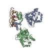 8r7yC M: map data used to model this data C: citing same article ( |
|---|---|
| Similar structure data |
-Links
 PDBj
PDBj


- Assembly
| Deposited unit | 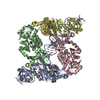
|
|---|---|
| 1 |
|
-Components
| #1: Protein | Mass: 38623.496 Da / Num. of mol.: 4 Source method: isolated from a genetically manipulated source Details: N-terminal amino-acid sequence GIDPFT remains a part of the protein upon TEV cleavage as a cloning artefact. Source: (gene. exp.) #2: DNA chain | | Mass: 13780.870 Da / Num. of mol.: 1 / Source method: obtained synthetically Details: Only 41bp were modelled into the electron density map (two base pairs at both ends are missing). Source: (synth.) #3: DNA chain | | Mass: 13931.952 Da / Num. of mol.: 1 / Source method: obtained synthetically Details: Only 41bp were modelled into the electron density map (two base pairs at both sides are missing). Source: (synth.) Has ligand of interest | N | Has protein modification | Y | |
|---|
-Experimental details
-Experiment
| Experiment | Method: ELECTRON MICROSCOPY |
|---|---|
| EM experiment | Aggregation state: PARTICLE / 3D reconstruction method: single particle reconstruction |
- Sample preparation
| Component | Name: A complex of Central glycolytic genes regulator (CggR) from Bacillus subtilis and the DNA operator (OLR). Type: COMPLEX Details: A complex of full-length CggR with its 45bp DNA operator. Entity ID: all / Source: RECOMBINANT | ||||||||||||||||||||
|---|---|---|---|---|---|---|---|---|---|---|---|---|---|---|---|---|---|---|---|---|---|
| Molecular weight | Experimental value: NO | ||||||||||||||||||||
| Source (natural) | Organism: | ||||||||||||||||||||
| Source (recombinant) | Organism: | ||||||||||||||||||||
| Buffer solution | pH: 7.5 Details: 20 mM Tris-HCl, pH 7.5, 100 mM NaCl and 0.02% (v/v) beta-mercaptoethanol | ||||||||||||||||||||
| Buffer component |
| ||||||||||||||||||||
| Specimen | Conc.: 0.86 mg/ml / Embedding applied: NO / Shadowing applied: NO / Staining applied: NO / Vitrification applied: YES | ||||||||||||||||||||
| Specimen support | Grid material: COPPER / Grid type: C-flat | ||||||||||||||||||||
| Vitrification | Instrument: FEI VITROBOT MARK IV / Cryogen name: ETHANE / Humidity: 100 % / Chamber temperature: 277 K |
- Electron microscopy imaging
| Experimental equipment |  Model: Titan Krios / Image courtesy: FEI Company |
|---|---|
| Microscopy | Model: FEI TITAN KRIOS |
| Electron gun | Electron source:  FIELD EMISSION GUN / Accelerating voltage: 300 kV / Illumination mode: FLOOD BEAM FIELD EMISSION GUN / Accelerating voltage: 300 kV / Illumination mode: FLOOD BEAM |
| Electron lens | Mode: BRIGHT FIELD / Nominal magnification: 165000 X / Nominal defocus max: 3000 nm / Nominal defocus min: 1500 nm / Cs: 2.7 mm / C2 aperture diameter: 30 µm |
| Specimen holder | Cryogen: NITROGEN / Specimen holder model: FEI TITAN KRIOS AUTOGRID HOLDER |
| Image recording | Electron dose: 40 e/Å2 / Detector mode: COUNTING / Film or detector model: GATAN K2 QUANTUM (4k x 4k) |
| EM imaging optics | Energyfilter name: GIF Quantum LS / Energyfilter slit width: 20 eV |
- Processing
| EM software |
| |||||||||||||||||||||
|---|---|---|---|---|---|---|---|---|---|---|---|---|---|---|---|---|---|---|---|---|---|---|
| CTF correction | Type: PHASE FLIPPING AND AMPLITUDE CORRECTION | |||||||||||||||||||||
| 3D reconstruction | Resolution: 4.4 Å / Resolution method: FSC 0.143 CUT-OFF / Num. of particles: 220146 / Algorithm: FOURIER SPACE / Symmetry type: POINT | |||||||||||||||||||||
| Atomic model building | B value: 24.32 / Protocol: RIGID BODY FIT / Space: REAL Details: Rigid body fitting of starting PDB models in Phenix and Coot was combined with manual rebuilding of certain model regions in Coot. | |||||||||||||||||||||
| Atomic model building |
|