 Movie
Movie Controller

[English] 日本語
 Yorodumi
- PDB-8r7y: Deoxyribonucleoside regulator DeoR in complex with the DNA operator -
+ Open data

- Basic information
| Entry | Database: PDB / ID: 8r7y | ||||||
|---|---|---|---|---|---|---|---|
| Title | Deoxyribonucleoside regulator DeoR in complex with the DNA operator | ||||||
 Components Components |
| ||||||
 Keywords Keywords | DNA BINDING PROTEIN / transcriptional repressor / protein-DNA complex / Bacillus subtilis | ||||||
| Function / homology |  Function and homology information Function and homology informationregulation of DNA-templated transcription initiation / cis-regulatory region sequence-specific DNA binding / carbohydrate binding / identical protein binding Similarity search - Function | ||||||
| Biological species |  | ||||||
| Method |  X-RAY DIFFRACTION / SYNCHROTRON / MOLECULAR REPLACEMENT / Resolution: 3.7 Å X-RAY DIFFRACTION / SYNCHROTRON / MOLECULAR REPLACEMENT / Resolution: 3.7 Å | ||||||
 Authors Authors | Pachl, P. / Soltysova, M. / Rezacova, P. | ||||||
| Funding support |  Czech Republic, 1items Czech Republic, 1items
| ||||||
 Citation Citation | Journal: Nucleic Acids Res / Year: 2024 Title: Structural characterization of two prototypical repressors of SorC family reveals tetrameric assemblies on DNA and mechanism of function. Authors: Markéta Šoltysová / Jana Škerlová / Petr Pachl / Karel Škubník / Milan Fábry / Irena Sieglová / Martina Farolfi / Irina Grishkovskaya / Michal Babiak / Jiří Nováček / Libor ...Authors: Markéta Šoltysová / Jana Škerlová / Petr Pachl / Karel Škubník / Milan Fábry / Irena Sieglová / Martina Farolfi / Irina Grishkovskaya / Michal Babiak / Jiří Nováček / Libor Krásný / Pavlína Řezáčová /  Abstract: The SorC family of transcriptional regulators plays a crucial role in controlling the carbohydrate metabolism and quorum sensing. We employed an integrative approach combining X-ray crystallography ...The SorC family of transcriptional regulators plays a crucial role in controlling the carbohydrate metabolism and quorum sensing. We employed an integrative approach combining X-ray crystallography and cryo-electron microscopy to investigate architecture and functional mechanism of two prototypical representatives of two sub-classes of the SorC family: DeoR and CggR from Bacillus subtilis. Despite possessing distinct DNA-binding domains, both proteins form similar tetrameric assemblies when bound to their respective DNA operators. Structural analysis elucidates the process by which the CggR-regulated gapA operon is derepressed through the action of two effectors: fructose-1,6-bisphosphate and newly confirmed dihydroxyacetone phosphate. Our findings provide the first comprehensive understanding of the DNA binding mechanism of the SorC-family proteins, shedding new light on their functional characteristics. | ||||||
| History |
|
- Structure visualization
| Structure viewer | Molecule: MolmilJmol/JSmol |
|---|
- Downloads & links
-Download
| PDBx/mmCIF format | 8r7y.cif.gz | 305 KB | Display | PDBx/mmCIF format |
|---|---|---|---|---|
| PDB format | pdb8r7y.ent.gz | 233.6 KB | Display | PDB format |
| PDBx/mmJSON format | 8r7y.json.gz | Tree view | PDBx/mmJSON format | |
| Others |  Other downloads Other downloads |
-Validation report
| Arichive directory | https://data.pdbj.org/pub/pdb/validation_reports/r7/8r7yftp://data.pdbj.org/pub/pdb/validation_reports/r7/8r7y | HTTPS FTP |
|---|
-Related structure data
-Links
 PDBj
PDBj


- Assembly
| Deposited unit | 
| ||||||||||||||||||||||||||||||||||||
|---|---|---|---|---|---|---|---|---|---|---|---|---|---|---|---|---|---|---|---|---|---|---|---|---|---|---|---|---|---|---|---|---|---|---|---|---|---|
| 1 | 
| ||||||||||||||||||||||||||||||||||||
| 2 | 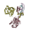
| ||||||||||||||||||||||||||||||||||||
| Unit cell |
| ||||||||||||||||||||||||||||||||||||
| Noncrystallographic symmetry (NCS) | NCS domain:
NCS domain segments: Beg auth comp-ID: THR / Beg label comp-ID: THR / End auth comp-ID: ASP / End label comp-ID: ASP / Auth asym-ID: A / Label asym-ID: A / Auth seq-ID: 1 - 312 / Label seq-ID: 6 - 317
NCS ensembles :
|
-Components
| #1: Protein | Mass: 35796.676 Da / Num. of mol.: 4 Source method: isolated from a genetically manipulated source Details: The N-terminal GIDPFT sequence is a cloning artefact. Source: (gene. exp.) Gene: deoR / Production host: #2: DNA chain | Mass: 5519.612 Da / Num. of mol.: 2 / Source method: obtained synthetically Source: (synth.) #3: DNA chain | Mass: 5506.630 Da / Num. of mol.: 2 / Source method: obtained synthetically Source: (synth.) #4: Water | ChemComp-HOH / |  Mass: 18.015 Da / Num. of mol.: 8 / Source method: isolated from a natural source / Formula: H2O Mass: 18.015 Da / Num. of mol.: 8 / Source method: isolated from a natural source / Formula: H2O |
|---|
-Experimental details
-Experiment
| Experiment | Method: X-RAY DIFFRACTION / Number of used crystals: 1 |
|---|
- Sample preparation
| Crystal | Density Matthews: 4.22 Å3/Da / Density % sol: 70.6 % / Description: Thin needle-shaped crystal. |
|---|---|
| Crystal grow | Temperature: 292 K / Method: vapor diffusion, sitting drop / pH: 6.3 Details: The volume ratio of the protein/DNA solution and precipitant solution - 1:2 Precipitant solution: 50 mM sodium cacodylate, pH 6.3, 75 mM CaCl2, 25 mM MgCl2, 3.5% (v/v) PEG 2K Crystal was ...Details: The volume ratio of the protein/DNA solution and precipitant solution - 1:2 Precipitant solution: 50 mM sodium cacodylate, pH 6.3, 75 mM CaCl2, 25 mM MgCl2, 3.5% (v/v) PEG 2K Crystal was cryo-protected by a quick soaking in 25% (v/v) glycerol. Temp details: Grown in an air-conditioned room. |
-Data collection
| Diffraction | Mean temperature: 100 K / Serial crystal experiment: N | ||||||||||||||||||||||||||||
|---|---|---|---|---|---|---|---|---|---|---|---|---|---|---|---|---|---|---|---|---|---|---|---|---|---|---|---|---|---|
| Diffraction source | Source: SYNCHROTRON / Site: BESSY  / Beamline: 14.1 / Wavelength: 0.9141 Å / Beamline: 14.1 / Wavelength: 0.9141 Å | ||||||||||||||||||||||||||||
| Detector | Type: DECTRIS PILATUS 6M / Detector: PIXEL / Date: Jan 31, 2018 | ||||||||||||||||||||||||||||
| Radiation | Protocol: SINGLE WAVELENGTH / Monochromatic (M) / Laue (L): M / Scattering type: x-ray | ||||||||||||||||||||||||||||
| Radiation wavelength | Wavelength: 0.9141 Å / Relative weight: 1 | ||||||||||||||||||||||||||||
| Reflection | Resolution: 3.6→50 Å / Num. obs: 32081 / % possible obs: 98.9 % / Redundancy: 13.1 % / Biso Wilson estimate: 122.8 Å2 / CC1/2: 0.993 / Rrim(I) all: 0.748 / Net I/σ(I): 4.86 | ||||||||||||||||||||||||||||
| Reflection shell |
|
- Processing
| Software |
| |||||||||||||||||||||||||||||||||||||||||||||||||||||||||||||||||||||||||||||||||||||||||||||||||||||||||||||||||||||||||||||||||||||||||||||||||||||||||||||||||||||||||||||||||||||||||||||||||||||||||||||||||||||||||||||||||||||||
|---|---|---|---|---|---|---|---|---|---|---|---|---|---|---|---|---|---|---|---|---|---|---|---|---|---|---|---|---|---|---|---|---|---|---|---|---|---|---|---|---|---|---|---|---|---|---|---|---|---|---|---|---|---|---|---|---|---|---|---|---|---|---|---|---|---|---|---|---|---|---|---|---|---|---|---|---|---|---|---|---|---|---|---|---|---|---|---|---|---|---|---|---|---|---|---|---|---|---|---|---|---|---|---|---|---|---|---|---|---|---|---|---|---|---|---|---|---|---|---|---|---|---|---|---|---|---|---|---|---|---|---|---|---|---|---|---|---|---|---|---|---|---|---|---|---|---|---|---|---|---|---|---|---|---|---|---|---|---|---|---|---|---|---|---|---|---|---|---|---|---|---|---|---|---|---|---|---|---|---|---|---|---|---|---|---|---|---|---|---|---|---|---|---|---|---|---|---|---|---|---|---|---|---|---|---|---|---|---|---|---|---|---|---|---|---|---|---|---|---|---|---|---|---|---|---|---|---|---|---|---|---|---|
| Refinement | Method to determine structure: MOLECULAR REPLACEMENT / Resolution: 3.7→48.924 Å / Cor.coef. Fo:Fc: 0.94 / Cor.coef. Fo:Fc free: 0.901 / SU B: 91.089 / SU ML: 1.116 / Cross valid method: FREE R-VALUE / ESU R Free: 0.795 Details: Hydrogens have been used if present in the input file
| |||||||||||||||||||||||||||||||||||||||||||||||||||||||||||||||||||||||||||||||||||||||||||||||||||||||||||||||||||||||||||||||||||||||||||||||||||||||||||||||||||||||||||||||||||||||||||||||||||||||||||||||||||||||||||||||||||||||
| Solvent computation | Ion probe radii: 0.8 Å / Shrinkage radii: 0.8 Å / VDW probe radii: 1.2 Å / Solvent model: MASK BULK SOLVENT | |||||||||||||||||||||||||||||||||||||||||||||||||||||||||||||||||||||||||||||||||||||||||||||||||||||||||||||||||||||||||||||||||||||||||||||||||||||||||||||||||||||||||||||||||||||||||||||||||||||||||||||||||||||||||||||||||||||||
| Displacement parameters | Biso mean: 172.198 Å2
| |||||||||||||||||||||||||||||||||||||||||||||||||||||||||||||||||||||||||||||||||||||||||||||||||||||||||||||||||||||||||||||||||||||||||||||||||||||||||||||||||||||||||||||||||||||||||||||||||||||||||||||||||||||||||||||||||||||||
| Refinement step | Cycle: LAST / Resolution: 3.7→48.924 Å
| |||||||||||||||||||||||||||||||||||||||||||||||||||||||||||||||||||||||||||||||||||||||||||||||||||||||||||||||||||||||||||||||||||||||||||||||||||||||||||||||||||||||||||||||||||||||||||||||||||||||||||||||||||||||||||||||||||||||
| Refine LS restraints |
| |||||||||||||||||||||||||||||||||||||||||||||||||||||||||||||||||||||||||||||||||||||||||||||||||||||||||||||||||||||||||||||||||||||||||||||||||||||||||||||||||||||||||||||||||||||||||||||||||||||||||||||||||||||||||||||||||||||||
| Refine LS restraints NCS | Auth asym-ID: A / Refine-ID: X-RAY DIFFRACTION / Type: medium positional; mediume thermal / Weight Biso : 2 / Weight position: 0.5
| |||||||||||||||||||||||||||||||||||||||||||||||||||||||||||||||||||||||||||||||||||||||||||||||||||||||||||||||||||||||||||||||||||||||||||||||||||||||||||||||||||||||||||||||||||||||||||||||||||||||||||||||||||||||||||||||||||||||
| LS refinement shell | Refine-ID: X-RAY DIFFRACTION / Total num. of bins used: 20
|
