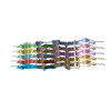
Movie
Movie Controller


+ Open data

- Basic information
| Entry | Database: PDB / ID: 8qpz | |||||||||
|---|---|---|---|---|---|---|---|---|---|---|
| Title | CryoEM structure of recombinant DeltaN7 alpha-synuclein in PBS | |||||||||
 Components Components | Alpha-synuclein | |||||||||
 Keywords Keywords | PROTEIN FIBRIL / synuclein / Parkinson's disease / neurodegeneration / amyloid / helical / fibril | |||||||||
| Function / homology |  Function and homology information Function and homology informationnegative regulation of mitochondrial electron transport, NADH to ubiquinone / negative regulation of dopamine uptake involved in synaptic transmission / negative regulation of norepinephrine uptake / response to desipramine / positive regulation of SNARE complex assembly / positive regulation of hydrogen peroxide catabolic process / supramolecular fiber / regulation of synaptic vesicle recycling / negative regulation of chaperone-mediated autophagy / regulation of reactive oxygen species biosynthetic process ...negative regulation of mitochondrial electron transport, NADH to ubiquinone / negative regulation of dopamine uptake involved in synaptic transmission / negative regulation of norepinephrine uptake / response to desipramine / positive regulation of SNARE complex assembly / positive regulation of hydrogen peroxide catabolic process / supramolecular fiber / regulation of synaptic vesicle recycling / negative regulation of chaperone-mediated autophagy / regulation of reactive oxygen species biosynthetic process / positive regulation of protein localization to cell periphery / negative regulation of exocytosis / dopamine biosynthetic process / dopamine uptake involved in synaptic transmission / negative regulation of dopamine metabolic process / response to iron(II) ion / negative regulation of platelet-derived growth factor receptor signaling pathway / SNARE complex assembly / negative regulation of thrombin-activated receptor signaling pathway / Lewy body / negative regulation of microtubule polymerization / synaptic vesicle priming / synaptic vesicle transport / regulation of norepinephrine uptake / transporter regulator activity / positive regulation of inositol phosphate biosynthetic process / protein kinase inhibitor activity / regulation of dopamine secretion / positive regulation of receptor recycling / cuprous ion binding / positive regulation of exocytosis / nuclear outer membrane / dynein complex binding / synaptic transmission, dopaminergic / synaptic vesicle exocytosis / response to magnesium ion / positive regulation of endocytosis / negative regulation of serotonin uptake / kinesin binding / cysteine-type endopeptidase inhibitor activity / regulation of presynapse assembly / synaptic vesicle endocytosis / alpha-tubulin binding / beta-tubulin binding / phospholipase binding / behavioral response to cocaine / cellular response to fibroblast growth factor stimulus / supramolecular fiber organization / response to type II interferon / cellular response to epinephrine stimulus / inclusion body / response to interleukin-1 / Hsp70 protein binding / positive regulation of release of sequestered calcium ion into cytosol / cellular response to copper ion / axon terminus / enzyme inhibitor activity / glutathione metabolic process / regulation of microtubule cytoskeleton organization / SNARE binding / protein tetramerization / protein sequestering activity / receptor internalization / phosphoprotein binding / microglial cell activation / tubulin binding / protein destabilization / ferrous iron binding / synapse organization / phospholipid binding / PKR-mediated signaling / tau protein binding / enzyme activator activity / positive regulation of inflammatory response / terminal bouton / actin cytoskeleton / synaptic vesicle membrane / growth cone / response to lipopolysaccharide / cellular response to oxidative stress / actin binding / histone binding / cell cortex / negative regulation of neuron apoptotic process / microtubule binding / amyloid fibril formation / mitochondrial outer membrane / lysosome / oxidoreductase activity / mitochondrial inner membrane / transcription cis-regulatory region binding / positive regulation of apoptotic process / ribosome / mitochondrial matrix / Amyloid fiber formation / copper ion binding / protein domain specific binding / axon / neuronal cell body / calcium ion binding Similarity search - Function | |||||||||
| Biological species |  Homo sapiens (human) Homo sapiens (human) | |||||||||
| Method | ELECTRON MICROSCOPY / helical reconstruction / cryo EM / Resolution: 2.5 Å | |||||||||
 Authors Authors | Thacker, D. / Wilkinson, M. / Dewison, K.M. / Ranson, N.A. / Brockwell, D.J. / Radford, S.E. | |||||||||
| Funding support |  United Kingdom, 2items United Kingdom, 2items
| |||||||||
 Citation Citation | Journal: Proc Natl Acad Sci U S A / Year: 2024 Title: Residues 2 to 7 of α-synuclein regulate amyloid formation via lipid-dependent and lipid-independent pathways. Authors: Katherine M Dewison / Benjamin Rowlinson / Jonathan M Machin / Joel A Crossley / Dev Thacker / Martin Wilkinson / Sabine M Ulamec / G Nasir Khan / Neil A Ranson / Patricija van Oosten-Hawle ...Authors: Katherine M Dewison / Benjamin Rowlinson / Jonathan M Machin / Joel A Crossley / Dev Thacker / Martin Wilkinson / Sabine M Ulamec / G Nasir Khan / Neil A Ranson / Patricija van Oosten-Hawle / David J Brockwell / Sheena E Radford /  Abstract: Amyloid formation by α-synuclein (αSyn) occurs in Parkinson's disease, multiple system atrophy, and dementia with Lewy bodies. Deciphering the residues that regulate αSyn amyloid fibril formation ...Amyloid formation by α-synuclein (αSyn) occurs in Parkinson's disease, multiple system atrophy, and dementia with Lewy bodies. Deciphering the residues that regulate αSyn amyloid fibril formation will not only provide mechanistic insight but may also reveal targets to prevent and treat disease. Previous investigations have identified several regions of αSyn to be important in the regulation of amyloid formation, including the non-amyloid-β component (NAC), P1 region (residues 36 to 42), and residues in the C-terminal domain. Recent studies have also indicated the importance of the N-terminal region of αSyn for both its physiological and pathological roles. Here, the role of residues 2 to 7 in the N-terminal region of αSyn is investigated in terms of their ability to regulate amyloid fibril formation in vitro and in vivo. Deletion of these residues (αSynΔN7) slows the rate of fibril formation in vitro and reduces the capacity of the protein to be recruited by wild-type (αSynWT) fibril seeds, despite cryo-EM showing a fibril structure consistent with those of full-length αSyn. Strikingly, fibril formation of αSynΔN7 is not induced by liposomes, despite the protein binding to liposomes with similar affinity to αSynWT. A model also showed that αSynΔN7::YFP forms few puncta and lacks motility and lifespan defects typified by expression of αSynWT::YFP. Together, the results demonstrate the involvement of residues 2 to 7 of αSyn in amyloid formation, revealing a target for the design of amyloid inhibitors that may leave the functional role of the protein in membrane binding unperturbed. | |||||||||
| History |
|
- Structure visualization
| Structure viewer | Molecule: MolmilJmol/JSmol |
|---|
- Downloads & links
-Download
| PDBx/mmCIF format | 8qpz.cif.gz | 109.5 KB | Display | PDBx/mmCIF format |
|---|---|---|---|---|
| PDB format | pdb8qpz.ent.gz | 79.5 KB | Display | PDB format |
| PDBx/mmJSON format | 8qpz.json.gz | Tree view | PDBx/mmJSON format | |
| Others |  Other downloads Other downloads |
-Validation report
| Arichive directory | https://data.pdbj.org/pub/pdb/validation_reports/qp/8qpzftp://data.pdbj.org/pub/pdb/validation_reports/qp/8qpz | HTTPS FTP |
|---|
-Related structure data
| Related structure data | 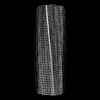 18570MC M: map data used to model this data C: citing same article ( |
|---|---|
| Similar structure data |
-Links
 PDBj
PDBj
- Assembly
| Deposited unit | 
|
|---|---|
| 1 |
|
-Components
| #1: Protein | Mass: 13797.286 Da / Num. of mol.: 12 / Mutation: deltaN7 (methionine kept for expression) Source method: isolated from a genetically manipulated source Details: DeltaN7, technically residues 2-7 are deleted as the N-terminal Methionine was required for bacterial expression. Source: (gene. exp.) Homo sapiens (human) / Gene: SNCA / Variant: DeltaN7 / Plasmid: pET23a / Production host:   Escherichia coli BL21(DE3) (bacteria) / References: UniProt: P37840 Escherichia coli BL21(DE3) (bacteria) / References: UniProt: P37840 |
|---|
-Experimental details
-Experiment
| Experiment | Method: ELECTRON MICROSCOPY |
|---|---|
| EM experiment | Aggregation state: FILAMENT / 3D reconstruction method: helical reconstruction |
- Sample preparation
| Component | Name: alpha-synuclein DeltaN7 amyloid fibrils / Type: COMPLEX / Entity ID: all / Source: RECOMBINANT |
|---|---|
| Molecular weight | Experimental value: NO |
| Source (natural) | Organism: Homo sapiens (human) |
| Source (recombinant) | Organism: Escherichia coli (E. coli) |
| Buffer solution | pH: 7.4 Details: 137 mM NaCl, 2.7 mM KCl, 8.1 mM Na2HPO4 and 1.5 mM KH2PO4; pH 7.4 |
| Specimen | Embedding applied: NO / Shadowing applied: NO / Staining applied: NO / Vitrification applied: YES |
| Specimen support | Grid material: COPPER / Grid mesh size: 300 divisions/in. |
| Vitrification | Instrument: FEI VITROBOT MARK IV / Cryogen name: ETHANE / Humidity: 90 % / Chamber temperature: 277 K |
- Electron microscopy imaging
| Experimental equipment |  Model: Titan Krios / Image courtesy: FEI Company |
|---|---|
| Microscopy | Model: FEI TITAN KRIOS |
| Electron gun | Electron source:  FIELD EMISSION GUN / Accelerating voltage: 300 kV / Illumination mode: FLOOD BEAM FIELD EMISSION GUN / Accelerating voltage: 300 kV / Illumination mode: FLOOD BEAM |
| Electron lens | Mode: BRIGHT FIELD / Nominal magnification: 130000 X / Nominal defocus max: 2600 nm / Nominal defocus min: 1400 nm / Cs: 2.7 mm / C2 aperture diameter: 50 µm / Alignment procedure: COMA FREE |
| Specimen holder | Specimen holder model: FEI TITAN KRIOS AUTOGRID HOLDER |
| Image recording | Electron dose: 45 e/Å2 / Film or detector model: FEI FALCON IV (4k x 4k) / Num. of grids imaged: 1 / Num. of real images: 5464 |
| EM imaging optics | Energyfilter name: TFS Selectris / Energyfilter slit width: 10 eV |
| Image scans | Width: 4096 / Height: 4096 |
- Processing
| EM software |
| ||||||||||||||||||||||||||||||||||||||||
|---|---|---|---|---|---|---|---|---|---|---|---|---|---|---|---|---|---|---|---|---|---|---|---|---|---|---|---|---|---|---|---|---|---|---|---|---|---|---|---|---|---|
| CTF correction | Type: PHASE FLIPPING AND AMPLITUDE CORRECTION | ||||||||||||||||||||||||||||||||||||||||
| Helical symmerty | Angular rotation/subunit: 179.43 ° / Axial rise/subunit: 2.44 Å / Axial symmetry: C1 | ||||||||||||||||||||||||||||||||||||||||
| Particle selection | Num. of particles selected: 315777 | ||||||||||||||||||||||||||||||||||||||||
| 3D reconstruction | Resolution: 2.5 Å / Resolution method: FSC 0.143 CUT-OFF / Num. of particles: 12348 / Symmetry type: HELICAL | ||||||||||||||||||||||||||||||||||||||||
| Atomic model building | B value: 106 / Protocol: RIGID BODY FIT / Space: REAL / Target criteria: cross-correlation coefficient Details: see methods, rigid body docked chain of PDB: 6osl, manual fitting in coot, real-space refine in phenix | ||||||||||||||||||||||||||||||||||||||||
| Atomic model building | PDB-ID: 6osl Pdb chain-ID: A / Accession code: 6osl / Chain residue range: 42-92 / Pdb chain residue range: 42-92 / Source name: PDB / Type: experimental model | ||||||||||||||||||||||||||||||||||||||||
| Refine LS restraints |
|