Journal: Proc Natl Acad Sci U S A / Year: 2024 Title: Residues 2 to 7 of α-synuclein regulate amyloid formation via lipid-dependent and lipid-independent pathways. Authors: Katherine M Dewison / Benjamin Rowlinson / Jonathan M Machin / Joel A Crossley / Dev Thacker / Martin Wilkinson / Sabine M Ulamec / G Nasir Khan / Neil A Ranson / Patricija van Oosten-Hawle ...Authors: Katherine M Dewison / Benjamin Rowlinson / Jonathan M Machin / Joel A Crossley / Dev Thacker / Martin Wilkinson / Sabine M Ulamec / G Nasir Khan / Neil A Ranson / Patricija van Oosten-Hawle / David J Brockwell / Sheena E Radford / Abstract: Amyloid formation by α-synuclein (αSyn) occurs in Parkinson's disease, multiple system atrophy, and dementia with Lewy bodies. Deciphering the residues that regulate αSyn amyloid fibril formation ...Amyloid formation by α-synuclein (αSyn) occurs in Parkinson's disease, multiple system atrophy, and dementia with Lewy bodies. Deciphering the residues that regulate αSyn amyloid fibril formation will not only provide mechanistic insight but may also reveal targets to prevent and treat disease. Previous investigations have identified several regions of αSyn to be important in the regulation of amyloid formation, including the non-amyloid-β component (NAC), P1 region (residues 36 to 42), and residues in the C-terminal domain. Recent studies have also indicated the importance of the N-terminal region of αSyn for both its physiological and pathological roles. Here, the role of residues 2 to 7 in the N-terminal region of αSyn is investigated in terms of their ability to regulate amyloid fibril formation in vitro and in vivo. Deletion of these residues (αSynΔN7) slows the rate of fibril formation in vitro and reduces the capacity of the protein to be recruited by wild-type (αSynWT) fibril seeds, despite cryo-EM showing a fibril structure consistent with those of full-length αSyn. Strikingly, fibril formation of αSynΔN7 is not induced by liposomes, despite the protein binding to liposomes with similar affinity to αSynWT. A model also showed that αSynΔN7::YFP forms few puncta and lacks motility and lifespan defects typified by expression of αSynWT::YFP. Together, the results demonstrate the involvement of residues 2 to 7 of αSyn in amyloid formation, revealing a target for the design of amyloid inhibitors that may leave the functional role of the protein in membrane binding unperturbed.
Name: Alpha-synuclein / type: protein_or_peptide / ID: 1 Details: DeltaN7, technically residues 2-7 are deleted as the N-terminal Methionine was required for bacterial expression. Number of copies: 12 / Enantiomer: LEVO
pH: 7.4 Details: 137 mM NaCl, 2.7 mM KCl, 8.1 mM Na2HPO4 and 1.5 mM KH2PO4; pH 7.4
Grid
Material: COPPER / Mesh: 300 / Support film - Material: CARBON / Support film - topology: LACEY / Pretreatment - Type: PLASMA CLEANING / Pretreatment - Time: 60 sec.
Vitrification
Cryogen name: ETHANE / Chamber humidity: 90 % / Chamber temperature: 277 K / Instrument: FEI VITROBOT MARK IV
-
Electron microscopy
Microscope
FEI TITAN KRIOS
Specialist optics
Energy filter - Name: TFS Selectris / Energy filter - Slit width: 10 eV
Image recording
Film or detector model: FEI FALCON IV (4k x 4k) / Digitization - Dimensions - Width: 4096 pixel / Digitization - Dimensions - Height: 4096 pixel / Number grids imaged: 1 / Number real images: 5464 / Average electron dose: 45.0 e/Å2
Electron beam
Acceleration voltage: 300 kV / Electron source: FIELD EMISSION GUN
In the structure databanks used in Yorodumi, some data are registered as the other names, "COVID-19 virus" and "2019-nCoV". Here are the details of the virus and the list of structure data.
Jan 31, 2019. EMDB accession codes are about to change! (news from PDBe EMDB page)
EMDB accession codes are about to change! (news from PDBe EMDB page)
The allocation of 4 digits for EMDB accession codes will soon come to an end. Whilst these codes will remain in use, new EMDB accession codes will include an additional digit and will expand incrementally as the available range of codes is exhausted. The current 4-digit format prefixed with “EMD-” (i.e. EMD-XXXX) will advance to a 5-digit format (i.e. EMD-XXXXX), and so on. It is currently estimated that the 4-digit codes will be depleted around Spring 2019, at which point the 5-digit format will come into force.
The EM Navigator/Yorodumi systems omit the EMD- prefix.
Related info.:Q: What is EMD? / ID/Accession-code notation in Yorodumi/EM Navigator
Yorodumi is a browser for structure data from EMDB, PDB, SASBDB, etc.
This page is also the successor to EM Navigator detail page, and also detail information page/front-end page for Omokage search.
The word "yorodu" (or yorozu) is an old Japanese word meaning "ten thousand". "mi" (miru) is to see.
Related info.:EMDB / PDB / SASBDB / Comparison of 3 databanks / Yorodumi Search / Aug 31, 2016. New EM Navigator & Yorodumi / Yorodumi Papers / Jmol/JSmol / Function and homology information / Changes in new EM Navigator and Yorodumi
 Movie
Movie Controller


 Open data
 Basic information
 Map data
 Sample
 Keywords
 Function and homology information
 Homo sapiens (human)
 Authors
 United Kingdom, 2 items
  Citation
 Structure visualization
 Downloads & links
 emd_18570.png
 http://ftp.pdbj.org/pub/emdb/structures/EMD-18570
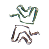
 Z (Sec.)
 Y (Row.)
 X (Col.)


 Sample components
 Processing
 Electron microscopy
 FIELD EMISSION GUN