[English] 日本語
Yorodumi
- PDB-8qjz: Crystal structure of E. coli LpxH in complex with lipid X -

+
Open data


ID or keywords:

Loading...

-
Basic information

Entry
Database: PDB / ID: 8qjz
TitleCrystal structure of E. coli LpxH in complex with lipid X
ComponentsUDP-2,3-diacylglucosamine hydrolase
KeywordsHYDROLASE / Lipid A biosynthesis pathway Endotoxin UDP-diacyl-glucosamine Lipid X Gram-negative bacteria Lipopolysaccharides HYDROLASE
Function / homology
Function and homology information


UDP-2,3-diacylglucosamine diphosphatase / UDP-2,3-diacylglucosamine hydrolase activity / lipid A biosynthetic process / extrinsic component of plasma membrane / manganese ion binding / cytoplasm
Similarity search - Function
UDP-2,3-diacylglucosamine hydrolase / UDP-2,3-diacylglucosamine hydrolase LpxH-like / Calcineurin-like phosphoesterase domain, ApaH type / Calcineurin-like phosphoesterase / Metallo-dependent phosphatase-like
Similarity search - Domain/homology
Chem-LP5 / : / UDP-2,3-diacylglucosamine hydrolase
Similarity search - Component
Biological speciesEscherichia coli (E. coli)
MethodX-RAY DIFFRACTION / SYNCHROTRON / MOLECULAR REPLACEMENT / Resolution: 1.35 Å
AuthorsSooriyaarachchi, S. / Bergfors, T. / Jones, T.A. / Mowbray, S.L.
Funding support Sweden, 1items
OrganizationGrant numberCountry
Swedish Research Council Sweden
CitationJournal: Proc.Natl.Acad.Sci.USA / Year: 2024
Title: Antibiotic class with potent in vivo activity targeting lipopolysaccharide synthesis in Gram-negative bacteria.
Authors: Huseby, D.L. / Cao, S. / Zamaratski, E. / Sooriyaarachchi, S. / Ahmad, S. / Bergfors, T. / Krasnova, L. / Pelss, J. / Ikaunieks, M. / Loza, E. / Katkevics, M. / Bobileva, O. / Cirule, H. / ...Authors: Huseby, D.L. / Cao, S. / Zamaratski, E. / Sooriyaarachchi, S. / Ahmad, S. / Bergfors, T. / Krasnova, L. / Pelss, J. / Ikaunieks, M. / Loza, E. / Katkevics, M. / Bobileva, O. / Cirule, H. / Gukalova, B. / Grinberga, S. / Backlund, M. / Simoff, I. / Leber, A.T. / Berruga-Fernandez, T. / Antonov, D. / Konda, V.R. / Lindstrom, S. / Olanders, G. / Brandt, P. / Baranczewski, P. / Vingsbo Lundberg, C. / Liepinsh, E. / Suna, E. / Jones, T.A. / Mowbray, S.L. / Hughes, D. / Karlen, A.
History
DepositionSep 14, 2023Deposition site: PDBE / Processing site: PDBE
Revision 1.0Apr 17, 2024Provider: repository / Type: Initial release

-
Structure visualization

Structure viewerMolecule:
MolmilJmol/JSmol

Downloads & links

-
Assembly

Deposited unit
A: UDP-2,3-diacylglucosamine hydrolase
hetero molecules


Theoretical massNumber of molelcules
Total (without water)28,6854
Polymers27,8631
Non-polymers8223
Water4,197233
1


  • Idetical with deposited unit
  • defined by author&software
TypeNameSymmetry operationNumber
identity operation1_555x,y,z1
Buried area1780 Å2
ΔGint-17 kcal/mol
Surface area11320 Å2
Unit cell
Length a, b, c (Å)123.827, 81.392, 33.127
Angle α, β, γ (deg.)90.00, 100.32, 90.00
Int Tables number5
Space group name H-MC121

-
Components

#1: Protein UDP-2,3-diacylglucosamine hydrolase


Mass: 27863.203 Da / Num. of mol.: 1
Source method: isolated from a genetically manipulated source
Source: (gene. exp.) Escherichia coli (E. coli) / Gene: lpxH / Plasmid: pET-26b(+) / Production host: Escherichia coli (E. coli) / Strain (production host): BL21(AI) / References: UniProt: A0A066QL39
#2: Chemical ChemComp-MN / MANGANESE (II) ION


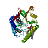
Mass: 54.938 Da / Num. of mol.: 2 / Source method: obtained synthetically / Formula: Mn
#3: Chemical ChemComp-LP5 / (R)-((2R,3S,4R,5R,6R)-3-HYDROXY-2-(HYDROXYMETHYL)-5-((R)-3-HYDROXYTETRADECANAMIDO)-6-(PHOSPHONOOXY)TETRAHYDRO-2H-PYRAN-4-YL) 3-HYDROXYTETRADECANOATE


Mass: 711.861 Da / Num. of mol.: 1 / Source method: obtained synthetically / Formula: C34H66NO12P
#4: Water ChemComp-HOH / water


Mass: 18.015 Da / Num. of mol.: 233 / Source method: isolated from a natural source / Formula: H2O
Has ligand of interestY

-
Experimental details

-
Experiment

ExperimentMethod: X-RAY DIFFRACTION / Number of used crystals: 1

-
Sample preparation

CrystalDensity Matthews: 2.97 Å3/Da / Density % sol: 58.57 % / Description: bipyramidal crystal
Crystal growTemperature: 293 K / Method: vapor diffusion / pH: 7.5
Details: Morpheus III (G7) 1.2 % Cholic acid derivatives mix 0.1 M Buffer System 2 7.5 50 % Precipitant Mix 3

-
Data collection

DiffractionMean temperature: 100 K / Serial crystal experiment: N
Diffraction sourceSource: SYNCHROTRON / Site: Diamond / Beamline: I04-1 / Wavelength: 0.91589 Å
DetectorType: DECTRIS PILATUS 6M-F / Detector: PIXEL / Date: Apr 11, 2019
Details: quasi-periodic elliptically polarizing undulator (qEPU)
RadiationProtocol: SINGLE WAVELENGTH / Monochromatic (M) / Laue (L): M / Scattering type: x-ray
Radiation wavelengthWavelength: 0.91589 Å / Relative weight: 1
ReflectionResolution: 1.35→67.6 Å / Num. obs: 70366 / % possible obs: 99 % / Redundancy: 6.1 % / CC1/2: 0.999 / Net I/σ(I): 11.1
Reflection shellResolution: 1.35→1.42 Å / Num. unique obs: 9911 / CC1/2: 0.827

-
Processing

Software
NameVersionClassification
REFMAC5.8.0352refinement
autoPROCdata reduction
SCALAdata scaling
PHASERphasing
RefinementMethod to determine structure: MOLECULAR REPLACEMENT / Resolution: 1.35→30 Å / Cor.coef. Fo:Fc: 0.971 / Cor.coef. Fo:Fc free: 0.97 / SU B: 0.916 / SU ML: 0.036 / Cross valid method: THROUGHOUT / ESU R: 0.046 / ESU R Free: 0.047 / Stereochemistry target values: MAXIMUM LIKELIHOOD / Details: HYDROGENS HAVE BEEN ADDED IN THE RIDING POSITIONS
RfactorNum. reflection% reflectionSelection details
Rfree0.18733 3468 4.9 %RANDOM
Rwork0.17146 ---
obs0.17224 66887 99.17 %-
Solvent computationIon probe radii: 0.8 Å / Shrinkage radii: 0.8 Å / VDW probe radii: 1.2 Å / Solvent model: MASK
Displacement parametersBiso mean: 16.599 Å2
Baniso -1Baniso -2Baniso -3
1-0.16 Å20 Å20 Å2
2---1.4 Å20 Å2
3---1.16 Å2
Refinement stepCycle: 1 / Resolution: 1.35→30 Å
ProteinNucleic acidLigandSolventTotal
Num. atoms1901 0 50 233 2184
Refine LS restraints
Refine-IDTypeDev idealDev ideal targetNumber
X-RAY DIFFRACTIONr_bond_refined_d0.0080.0122011
X-RAY DIFFRACTIONr_bond_other_d0.0010.0161819
X-RAY DIFFRACTIONr_angle_refined_deg1.2811.6522707
X-RAY DIFFRACTIONr_angle_other_deg0.4651.5734216
X-RAY DIFFRACTIONr_dihedral_angle_1_deg5.7825239
X-RAY DIFFRACTIONr_dihedral_angle_2_deg7.337515
X-RAY DIFFRACTIONr_dihedral_angle_3_deg11.81710328
X-RAY DIFFRACTIONr_dihedral_angle_4_deg
X-RAY DIFFRACTIONr_chiral_restr0.0650.2303
X-RAY DIFFRACTIONr_gen_planes_refined0.0060.022233
X-RAY DIFFRACTIONr_gen_planes_other0.0010.02407
X-RAY DIFFRACTIONr_nbd_refined
X-RAY DIFFRACTIONr_nbd_other
X-RAY DIFFRACTIONr_nbtor_refined
X-RAY DIFFRACTIONr_nbtor_other
X-RAY DIFFRACTIONr_xyhbond_nbd_refined
X-RAY DIFFRACTIONr_xyhbond_nbd_other
X-RAY DIFFRACTIONr_metal_ion_refined
X-RAY DIFFRACTIONr_metal_ion_other
X-RAY DIFFRACTIONr_symmetry_vdw_refined
X-RAY DIFFRACTIONr_symmetry_vdw_other
X-RAY DIFFRACTIONr_symmetry_hbond_refined
X-RAY DIFFRACTIONr_symmetry_hbond_other
X-RAY DIFFRACTIONr_symmetry_metal_ion_refined
X-RAY DIFFRACTIONr_symmetry_metal_ion_other
X-RAY DIFFRACTIONr_mcbond_it1.191.685962
X-RAY DIFFRACTIONr_mcbond_other1.1731.684962
X-RAY DIFFRACTIONr_mcangle_it1.8372.5281199
X-RAY DIFFRACTIONr_mcangle_other1.8372.5291200
X-RAY DIFFRACTIONr_scbond_it1.9681.9441049
X-RAY DIFFRACTIONr_scbond_other1.9451.9111000
X-RAY DIFFRACTIONr_scangle_it
X-RAY DIFFRACTIONr_scangle_other2.9332.781449
X-RAY DIFFRACTIONr_long_range_B_refined4.32725.682229
X-RAY DIFFRACTIONr_long_range_B_other4.13922.1232147
X-RAY DIFFRACTIONr_rigid_bond_restr
X-RAY DIFFRACTIONr_sphericity_free
X-RAY DIFFRACTIONr_sphericity_bonded
LS refinement shellResolution: 1.35→1.384 Å
RfactorNum. reflection% reflection
Rfree0.297 255 -
Rwork0.282 4713 -
obs--95.04 %

+
About Yorodumi

-
News

-
Feb 9, 2022. New format data for meta-information of EMDB entries

New format data for meta-information of EMDB entries

  • Version 3 of the EMDB header file is now the official format.
  • The previous official version 1.9 will be removed from the archive.

Related info.:EMDB header

External links:wwPDB to switch to version 3 of the EMDB data model

-
Aug 12, 2020. Covid-19 info

Covid-19 info

URL: https://pdbjlvh1.pdbj.org/emnavi/covid19.php

New page: Covid-19 featured information page in EM Navigator.

Related info.:Covid-19 info / Mar 5, 2020. Novel coronavirus structure data

+
Mar 5, 2020. Novel coronavirus structure data

Novel coronavirus structure data

Related info.:Yorodumi Speices / Aug 12, 2020. Covid-19 info

External links:COVID-19 featured content - PDBj / Molecule of the Month (242):Coronavirus Proteases

+
Jan 31, 2019. EMDB accession codes are about to change! (news from PDBe EMDB page)

EMDB accession codes are about to change! (news from PDBe EMDB page)

  • The allocation of 4 digits for EMDB accession codes will soon come to an end. Whilst these codes will remain in use, new EMDB accession codes will include an additional digit and will expand incrementally as the available range of codes is exhausted. The current 4-digit format prefixed with “EMD-” (i.e. EMD-XXXX) will advance to a 5-digit format (i.e. EMD-XXXXX), and so on. It is currently estimated that the 4-digit codes will be depleted around Spring 2019, at which point the 5-digit format will come into force.
  • The EM Navigator/Yorodumi systems omit the EMD- prefix.

Related info.:Q: What is EMD? / ID/Accession-code notation in Yorodumi/EM Navigator

External links:EMDB Accession Codes are Changing Soon! / Contact to PDBj

+
Jul 12, 2017. Major update of PDB

Major update of PDB

  • wwPDB released updated PDB data conforming to the new PDBx/mmCIF dictionary.
  • This is a major update changing the version number from 4 to 5, and with Remediation, in which all the entries are updated.
  • In this update, many items about electron microscopy experimental information are reorganized (e.g. em_software).
  • Now, EM Navigator and Yorodumi are based on the updated data.

External links:wwPDB Remediation / Enriched Model Files Conforming to OneDep Data Standards Now Available in the PDB FTP Archive

-
Yorodumi

Thousand views of thousand structures

  • Yorodumi is a browser for structure data from EMDB, PDB, SASBDB, etc.
  • This page is also the successor to EM Navigator detail page, and also detail information page/front-end page for Omokage search.
  • The word "yorodu" (or yorozu) is an old Japanese word meaning "ten thousand". "mi" (miru) is to see.

Related info.:EMDB / PDB / SASBDB / Comparison of 3 databanks / Yorodumi Search / Aug 31, 2016. New EM Navigator & Yorodumi / Yorodumi Papers / Jmol/JSmol / Function and homology information / Changes in new EM Navigator and Yorodumi

Read more