 Movie
Movie Controller

[English] 日本語
 Yorodumi
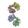
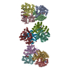
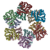
- PDB-8q50: Nitrogenase Fe protein from Methanothermococcus thermolithotrophi... -
+ Open data

- Basic information
| Entry | Database: PDB / ID: 8q50 | |||||||||
|---|---|---|---|---|---|---|---|---|---|---|
| Title | Nitrogenase Fe protein from Methanothermococcus thermolithotrophicus, tetragonal crystalline form at 1.91-A resolution | |||||||||
 Components Components | Nitrogenase iron protein 1 | |||||||||
 Keywords Keywords | ELECTRON TRANSPORT / nitrogenase / electron donor / ATPase / [4Fe-4S]-cluster / electron transfer / conformational changes / methanogenic archaea / oxidoreductase / O2-sensitivity / N2-fixation | |||||||||
| Function / homology |  Function and homology information Function and homology informationnitrogenase / nitrogenase activity / nitrogen fixation / 4 iron, 4 sulfur cluster binding / ATP binding / metal ion binding Similarity search - Function | |||||||||
| Biological species |  Methanothermococcus thermolithotrophicus DSM 2095 (archaea) Methanothermococcus thermolithotrophicus DSM 2095 (archaea) | |||||||||
| Method |  X-RAY DIFFRACTION / SYNCHROTRON / MOLECULAR REPLACEMENT / Resolution: 1.91 Å X-RAY DIFFRACTION / SYNCHROTRON / MOLECULAR REPLACEMENT / Resolution: 1.91 Å | |||||||||
 Authors Authors | Maslac, N. / Wagner, T. | |||||||||
| Funding support |  Germany, Germany,  Switzerland, 2items Switzerland, 2items
| |||||||||
 Citation Citation | Journal: Febs J. / Year: 2024 Title: Structural comparison of (hyper-)thermophilic nitrogenase reductases from three marine Methanococcales. Authors: Maslac, N. / Cadoux, C. / Bolte, P. / Murken, F. / Gu, W. / Milton, R.D. / Wagner, T. | |||||||||
| History |
|
- Structure visualization
| Structure viewer | Molecule: MolmilJmol/JSmol |
|---|
- Downloads & links
-Download
| PDBx/mmCIF format | 8q50.cif.gz | 126.3 KB | Display | PDBx/mmCIF format |
|---|---|---|---|---|
| PDB format | pdb8q50.ent.gz | 97.6 KB | Display | PDB format |
| PDBx/mmJSON format | 8q50.json.gz | Tree view | PDBx/mmJSON format | |
| Others |  Other downloads Other downloads |
-Validation report
| Arichive directory | https://data.pdbj.org/pub/pdb/validation_reports/q5/8q50ftp://data.pdbj.org/pub/pdb/validation_reports/q5/8q50 | HTTPS FTP |
|---|
-Related structure data

-Links
 PDBj
PDBj


- Assembly
| Deposited unit | 
| |||||||||
|---|---|---|---|---|---|---|---|---|---|---|
| 1 | 
| |||||||||
| Unit cell |
| |||||||||
| Components on special symmetry positions |
|
-Components
-Protein , 1 types, 1 molecules A
| #1: Protein | Mass: 31271.863 Da / Num. of mol.: 1 / Source method: isolated from a natural source Source: (natural) Methanothermococcus thermolithotrophicus DSM 2095 (archaea)Cell line: / / Organ: / / Plasmid details: / / Variant: / / Strain: SN-1 / Tissue: / / References: UniProt: P25767 |
|---|
-Non-polymers , 5 types, 133 molecules 


| #2: Chemical | ChemComp-CA /  Mass: 40.078 Da / Num. of mol.: 1 / Source method: obtained synthetically / Formula: Ca Mass: 40.078 Da / Num. of mol.: 1 / Source method: obtained synthetically / Formula: Ca | ||||
|---|---|---|---|---|---|
| #3: Chemical | ChemComp-FES /  Mass: 175.820 Da / Num. of mol.: 1 / Source method: obtained synthetically / Formula: Fe2S2 / Feature type: SUBJECT OF INVESTIGATION Mass: 175.820 Da / Num. of mol.: 1 / Source method: obtained synthetically / Formula: Fe2S2 / Feature type: SUBJECT OF INVESTIGATION | ||||
| #4: Chemical |  Mass: 92.094 Da / Num. of mol.: 2 / Source method: obtained synthetically / Formula: C3H8O3 Mass: 92.094 Da / Num. of mol.: 2 / Source method: obtained synthetically / Formula: C3H8O3#5: Chemical |  Mass: 59.044 Da / Num. of mol.: 3 / Source method: isolated from a natural source / Formula: C2H3O2 Mass: 59.044 Da / Num. of mol.: 3 / Source method: isolated from a natural source / Formula: C2H3O2#6: Water | ChemComp-HOH / | Mass: 18.015 Da / Num. of mol.: 126 / Source method: isolated from a natural source / Formula: H2O |
-Details
| Has ligand of interest | Y |
|---|
-Experimental details
-Experiment
| Experiment | Method: X-RAY DIFFRACTION / Number of used crystals: 1 |
|---|
- Sample preparation
| Crystal | Density Matthews: 2.39 Å3/Da / Density % sol: 48.55 % Description: The crystal was in a shape of an orthorhombic rod and brown. |
|---|---|
| Crystal grow | Temperature: 293.15 K / Method: vapor diffusion, sitting drop / pH: 4.6 Details: The purified protein was crystallised at a final concentration of 6 mg/ml by spotting 0.5 ul of crystallisation solution with 0.5 ul of protein sample. Crystallization was done anaerobically ...Details: The purified protein was crystallised at a final concentration of 6 mg/ml by spotting 0.5 ul of crystallisation solution with 0.5 ul of protein sample. Crystallization was done anaerobically in a Coy tent containing N2/H2 (97:3%) atmosphere. The screening was done at 20 degrees Celsius on 96-Well MRC 2-drop polystyrene (SWISSCI) plates containing 90 ul of crystallization solution. The crystallisation solution in which crystals were obtained contained 30 % v/v 2-methyl-2,4-pentanediol, 100 mM sodium acetate, pH 4.6 and 20 mM calcium chloride. |
-Data collection
| Diffraction | Mean temperature: 100 K / Serial crystal experiment: N |
|---|---|
| Diffraction source | Source: SYNCHROTRON / Site: SOLEIL  / Beamline: PROXIMA 1 / Wavelength: 1.74013 Å / Beamline: PROXIMA 1 / Wavelength: 1.74013 Å |
| Detector | Type: DECTRIS EIGER X 16M / Detector: PIXEL / Date: Nov 22, 2019 |
| Radiation | Protocol: SINGLE WAVELENGTH / Monochromatic (M) / Laue (L): M / Scattering type: x-ray |
| Radiation wavelength | Wavelength: 1.74013 Å / Relative weight: 1 |
| Reflection | Resolution: 1.91→52.01 Å / Num. obs: 24200 / % possible obs: 100 % / Redundancy: 41.2 % / CC1/2: 0.997 / Rmerge(I) obs: 0.159 / Rpim(I) all: 0.025 / Rrim(I) all: 0.161 / Net I/σ(I): 15.9 |
| Reflection shell | Resolution: 1.91→1.94 Å / Redundancy: 23.9 % / Rmerge(I) obs: 3.12 / Mean I/σ(I) obs: 1.4 / Num. unique obs: 1183 / CC1/2: 0.616 / Rpim(I) all: 0.644 / Rrim(I) all: 3.19 / % possible all: 100 |
- Processing
| Software |
| |||||||||||||||||||||||||||||||||||||||||||||||||||||||||||||||||||||||||||||||||||||||||||||||||||||||||||||||||||||||||||||||||||||||||||||||||||||||||||||||||||||||||||||||||||||||||||||||||||||||||||||||||||||||||||||||||||||||||||||||||||||||||||||||||||||||||||||||||||||||||||||||||||||||||||||||||||||||||||||||||||||
|---|---|---|---|---|---|---|---|---|---|---|---|---|---|---|---|---|---|---|---|---|---|---|---|---|---|---|---|---|---|---|---|---|---|---|---|---|---|---|---|---|---|---|---|---|---|---|---|---|---|---|---|---|---|---|---|---|---|---|---|---|---|---|---|---|---|---|---|---|---|---|---|---|---|---|---|---|---|---|---|---|---|---|---|---|---|---|---|---|---|---|---|---|---|---|---|---|---|---|---|---|---|---|---|---|---|---|---|---|---|---|---|---|---|---|---|---|---|---|---|---|---|---|---|---|---|---|---|---|---|---|---|---|---|---|---|---|---|---|---|---|---|---|---|---|---|---|---|---|---|---|---|---|---|---|---|---|---|---|---|---|---|---|---|---|---|---|---|---|---|---|---|---|---|---|---|---|---|---|---|---|---|---|---|---|---|---|---|---|---|---|---|---|---|---|---|---|---|---|---|---|---|---|---|---|---|---|---|---|---|---|---|---|---|---|---|---|---|---|---|---|---|---|---|---|---|---|---|---|---|---|---|---|---|---|---|---|---|---|---|---|---|---|---|---|---|---|---|---|---|---|---|---|---|---|---|---|---|---|---|---|---|---|---|---|---|---|---|---|---|---|---|---|---|---|---|---|---|---|---|---|---|---|---|---|---|---|---|---|---|---|---|---|---|---|---|---|---|---|---|---|---|---|---|---|---|---|---|---|---|---|---|---|---|---|---|---|---|---|---|---|---|---|---|---|---|---|
| Refinement | Method to determine structure: MOLECULAR REPLACEMENT / Resolution: 1.91→52.01 Å / SU ML: 0.25 / Cross valid method: FREE R-VALUE / σ(F): 1.35 / Phase error: 22.84 / Stereochemistry target values: ML Details: Translation-libration screw was used during refinement. The model was refined with riding hydrogens that were omitted in the final deposited model
| |||||||||||||||||||||||||||||||||||||||||||||||||||||||||||||||||||||||||||||||||||||||||||||||||||||||||||||||||||||||||||||||||||||||||||||||||||||||||||||||||||||||||||||||||||||||||||||||||||||||||||||||||||||||||||||||||||||||||||||||||||||||||||||||||||||||||||||||||||||||||||||||||||||||||||||||||||||||||||||||||||||
| Solvent computation | Shrinkage radii: 0.9 Å / VDW probe radii: 1.11 Å / Solvent model: FLAT BULK SOLVENT MODEL | |||||||||||||||||||||||||||||||||||||||||||||||||||||||||||||||||||||||||||||||||||||||||||||||||||||||||||||||||||||||||||||||||||||||||||||||||||||||||||||||||||||||||||||||||||||||||||||||||||||||||||||||||||||||||||||||||||||||||||||||||||||||||||||||||||||||||||||||||||||||||||||||||||||||||||||||||||||||||||||||||||||
| Displacement parameters | Biso mean: 46.31 Å2 | |||||||||||||||||||||||||||||||||||||||||||||||||||||||||||||||||||||||||||||||||||||||||||||||||||||||||||||||||||||||||||||||||||||||||||||||||||||||||||||||||||||||||||||||||||||||||||||||||||||||||||||||||||||||||||||||||||||||||||||||||||||||||||||||||||||||||||||||||||||||||||||||||||||||||||||||||||||||||||||||||||||
| Refinement step | Cycle: LAST / Resolution: 1.91→52.01 Å
| |||||||||||||||||||||||||||||||||||||||||||||||||||||||||||||||||||||||||||||||||||||||||||||||||||||||||||||||||||||||||||||||||||||||||||||||||||||||||||||||||||||||||||||||||||||||||||||||||||||||||||||||||||||||||||||||||||||||||||||||||||||||||||||||||||||||||||||||||||||||||||||||||||||||||||||||||||||||||||||||||||||
| Refine LS restraints |
| |||||||||||||||||||||||||||||||||||||||||||||||||||||||||||||||||||||||||||||||||||||||||||||||||||||||||||||||||||||||||||||||||||||||||||||||||||||||||||||||||||||||||||||||||||||||||||||||||||||||||||||||||||||||||||||||||||||||||||||||||||||||||||||||||||||||||||||||||||||||||||||||||||||||||||||||||||||||||||||||||||||
| LS refinement shell |
| |||||||||||||||||||||||||||||||||||||||||||||||||||||||||||||||||||||||||||||||||||||||||||||||||||||||||||||||||||||||||||||||||||||||||||||||||||||||||||||||||||||||||||||||||||||||||||||||||||||||||||||||||||||||||||||||||||||||||||||||||||||||||||||||||||||||||||||||||||||||||||||||||||||||||||||||||||||||||||||||||||||
| Refinement TLS params. | Method: refined / Refine-ID: X-RAY DIFFRACTION
| |||||||||||||||||||||||||||||||||||||||||||||||||||||||||||||||||||||||||||||||||||||||||||||||||||||||||||||||||||||||||||||||||||||||||||||||||||||||||||||||||||||||||||||||||||||||||||||||||||||||||||||||||||||||||||||||||||||||||||||||||||||||||||||||||||||||||||||||||||||||||||||||||||||||||||||||||||||||||||||||||||||
| Refinement TLS group |
|
