 Movie
Movie Controller

[English] 日本語
 Yorodumi
- PDB-8pdt: Crystal structure of Paradendryphiella salina PL7C alginate lyase... -
+ Open data

- Basic information
| Entry | Database: PDB / ID: 8pdt | ||||||
|---|---|---|---|---|---|---|---|
| Title | Crystal structure of Paradendryphiella salina PL7C alginate lyase soaked with dimannuronic acid | ||||||
 Components Components | Alginate lyase | ||||||
 Keywords Keywords | LYASE / Alginate lyase / complex / beta-jelly roll | ||||||
| Function / homology | Alginate lyase 2 / Alginate lyase / Concanavalin A-like lectin/glucanase domain superfamily / lyase activity / Alginate lyase Function and homology information Function and homology information | ||||||
| Biological species |  Paradendryphiella salina (fungus) Paradendryphiella salina (fungus) | ||||||
| Method |  X-RAY DIFFRACTION / SYNCHROTRON / MOLECULAR REPLACEMENT / Resolution: 1.09 Å X-RAY DIFFRACTION / SYNCHROTRON / MOLECULAR REPLACEMENT / Resolution: 1.09 Å | ||||||
 Authors Authors | Wilknes, C. | ||||||
| Funding support | 1items
| ||||||
 Citation Citation | Journal: Nat Commun / Year: 2025 Title: Unraveling the molecular mechanism of polysaccharide lyases for efficient alginate degradation. Authors: Rivas-Fernandez, J.P. / Vuillemin, M. / Pilgaard, B. / Klau, L.J. / Fredslund, F. / Lund-Hanssen, C. / Welner, D.H. / Meyer, A.S. / Morth, J.P. / Meilleur, F. / Aachmann, F.L. / Rovira, C. / Wilkens, C. | ||||||
| History |
|
- Structure visualization
| Structure viewer | Molecule: MolmilJmol/JSmol |
|---|
- Downloads & links
-Download
| PDBx/mmCIF format | 8pdt.cif.gz | 196.4 KB | Display | PDBx/mmCIF format |
|---|---|---|---|---|
| PDB format | pdb8pdt.ent.gz | 130 KB | Display | PDB format |
| PDBx/mmJSON format | 8pdt.json.gz | Tree view | PDBx/mmJSON format | |
| Others |  Other downloads Other downloads |
-Validation report
| Summary document | 8pdt_validation.pdf.gz | 1.2 MB | Display | wwPDB validaton report |
|---|---|---|---|---|
| Full document | 8pdt_full_validation.pdf.gz | 1.2 MB | Display | |
| Data in XML | 8pdt_validation.xml.gz | 14.1 KB | Display | |
| Data in CIF | 8pdt_validation.cif.gz | 21.9 KB | Display | |
| Arichive directory | https://data.pdbj.org/pub/pdb/validation_reports/pd/8pdtftp://data.pdbj.org/pub/pdb/validation_reports/pd/8pdt | HTTPS FTP |
-Related structure data
| Related structure data |  6ywfC  7nczC  7nl3C  7nm6C  7nppC  7ny3C  7o6hC  7oofC  7oryC  7p25C  7p90C  7pbfC  8bjoC  8bxzC  8c0mC  8c3xC  8p6oC  8pc3C  8pc8C  8pcxC  8pedC  8qizC  8qliC  8qmjC  8r43C  8rbiC  9g98C  9g99C C: citing same article ( |
|---|---|
| Similar structure data |
-Links
 PDBj
PDBj- Assembly
| Deposited unit | 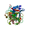
| ||||||||||||
|---|---|---|---|---|---|---|---|---|---|---|---|---|---|
| 1 |
| ||||||||||||
| Unit cell |
|
-Components
| #1: Protein | Mass: 25544.244 Da / Num. of mol.: 1 Source method: isolated from a genetically manipulated source Source: (gene. exp.) Paradendryphiella salina (fungus) / Gene: PsAlg7C / Production host: Komagataella pastoris (fungus) / References: UniProt: A0A7I9C8Z1 |
|---|---|
| #2: Polysaccharide | beta-D-mannopyranuronic acid-(1-4)-alpha-D-mannopyranuronic acid Source method: isolated from a genetically manipulated source |
| #3: Polysaccharide | beta-D-mannopyranuronic acid-(1-4)-alpha-L-gulopyranuronic acid Source method: isolated from a genetically manipulated source |
| #4: Water | ChemComp-HOH /  Mass: 18.015 Da / Num. of mol.: 306 / Source method: isolated from a natural source / Formula: H2O Mass: 18.015 Da / Num. of mol.: 306 / Source method: isolated from a natural source / Formula: H2O |
| Has ligand of interest | Y |
| Has protein modification | N |
-Experimental details
-Experiment
| Experiment | Method: X-RAY DIFFRACTION / Number of used crystals: 1 |
|---|
- Sample preparation
| Crystal | Density Matthews: 2.18 Å3/Da / Density % sol: 43.45 % |
|---|---|
| Crystal grow | Temperature: 293 K / Method: vapor diffusion, sitting drop / Details: 30% PEG3350, 0.1M Bis-Tris pH 5.5, 0.3M NaCl |
-Data collection
| Diffraction | Mean temperature: 100 K / Serial crystal experiment: N |
|---|---|
| Diffraction source | Source: SYNCHROTRON / Site: MAX IV  / Beamline: BioMAX / Wavelength: 0.799 Å / Beamline: BioMAX / Wavelength: 0.799 Å |
| Detector | Type: DECTRIS EIGER X 16M / Detector: PIXEL / Date: Aug 22, 2020 |
| Radiation | Protocol: SINGLE WAVELENGTH / Monochromatic (M) / Laue (L): M / Scattering type: x-ray |
| Radiation wavelength | Wavelength: 0.799 Å / Relative weight: 1 |
| Reflection | Resolution: 1.09→44.88 Å / Num. obs: 92201 / % possible obs: 98.4 % / Redundancy: 7.16 % / Biso Wilson estimate: 13.33 Å2 / CC1/2: 1 / Net I/σ(I): 7.77 |
| Reflection shell | Resolution: 1.09→1.16 Å / Num. unique obs: 14459 / CC1/2: 0.46 |
- Processing
| Software |
| ||||||||||||||||||||||||||||||||||||||||||||||||||||||||||||||||||||||||||||||||||||||||||||||||||||||||||||||||
|---|---|---|---|---|---|---|---|---|---|---|---|---|---|---|---|---|---|---|---|---|---|---|---|---|---|---|---|---|---|---|---|---|---|---|---|---|---|---|---|---|---|---|---|---|---|---|---|---|---|---|---|---|---|---|---|---|---|---|---|---|---|---|---|---|---|---|---|---|---|---|---|---|---|---|---|---|---|---|---|---|---|---|---|---|---|---|---|---|---|---|---|---|---|---|---|---|---|---|---|---|---|---|---|---|---|---|---|---|---|---|---|---|---|
| Refinement | Method to determine structure: MOLECULAR REPLACEMENT / Resolution: 1.09→37.77 Å / SU ML: 0.1232 / Cross valid method: FREE R-VALUE / σ(F): 1.35 / Phase error: 19.71 Stereochemistry target values: GeoStd + Monomer Library + CDL v1.2
| ||||||||||||||||||||||||||||||||||||||||||||||||||||||||||||||||||||||||||||||||||||||||||||||||||||||||||||||||
| Solvent computation | Shrinkage radii: 0.9 Å / VDW probe radii: 1.11 Å / Solvent model: FLAT BULK SOLVENT MODEL | ||||||||||||||||||||||||||||||||||||||||||||||||||||||||||||||||||||||||||||||||||||||||||||||||||||||||||||||||
| Displacement parameters | Biso mean: 18.07 Å2 | ||||||||||||||||||||||||||||||||||||||||||||||||||||||||||||||||||||||||||||||||||||||||||||||||||||||||||||||||
| Refinement step | Cycle: LAST / Resolution: 1.09→37.77 Å
| ||||||||||||||||||||||||||||||||||||||||||||||||||||||||||||||||||||||||||||||||||||||||||||||||||||||||||||||||
| Refine LS restraints |
| ||||||||||||||||||||||||||||||||||||||||||||||||||||||||||||||||||||||||||||||||||||||||||||||||||||||||||||||||
| LS refinement shell |
|
