 Movie
Movie Controller

[English] 日本語
 Yorodumi
- PDB-8pbw: Histidine-containing phosphotransfer protein from Chaetomium ther... -
+ Open data

- Basic information
| Entry | Database: PDB / ID: 8pbw | ||||||
|---|---|---|---|---|---|---|---|
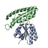
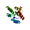
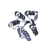
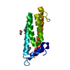
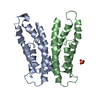
| Title | Histidine-containing phosphotransfer protein from Chaetomium thermophilum | ||||||
 Components Components | HPt domain-containing protein | ||||||
 Keywords Keywords | TRANSFERASE / Histidine-containing phosphotransferase / Fungal / Phosphorelay | ||||||
| Function / homology |  Function and homology information Function and homology informationprotein histidine kinase binding / histidine phosphotransfer kinase activity / phosphorelay signal transduction system / nucleus / cytoplasm Similarity search - Function | ||||||
| Biological species |  Thermochaetoides thermophila (fungus) Thermochaetoides thermophila (fungus) | ||||||
| Method |  X-RAY DIFFRACTION / SYNCHROTRON / MOLECULAR REPLACEMENT / Resolution: 2.4 Å X-RAY DIFFRACTION / SYNCHROTRON / MOLECULAR REPLACEMENT / Resolution: 2.4 Å | ||||||
 Authors Authors | Paredes-Martinez, F. / Casino, P. | ||||||
| Funding support |  Spain, 1items Spain, 1items
| ||||||
 Citation Citation | Journal: Commun Biol / Year: 2024 Title: Structural and functional insights underlying recognition of histidine phosphotransfer protein in fungal phosphorelay systems. Authors: Paredes-Martinez, F. / Eixeres, L. / Zamora-Caballero, S. / Casino, P. | ||||||
| History |
|
- Structure visualization
| Structure viewer | Molecule: MolmilJmol/JSmol |
|---|
- Downloads & links
-Download
| PDBx/mmCIF format | 8pbw.cif.gz | 113.8 KB | Display | PDBx/mmCIF format |
|---|---|---|---|---|
| PDB format | pdb8pbw.ent.gz | 87 KB | Display | PDB format |
| PDBx/mmJSON format | 8pbw.json.gz | Tree view | PDBx/mmJSON format | |
| Others |  Other downloads Other downloads |
-Validation report
| Arichive directory | https://data.pdbj.org/pub/pdb/validation_reports/pb/8pbwftp://data.pdbj.org/pub/pdb/validation_reports/pb/8pbw | HTTPS FTP |
|---|
-Related structure data
-Links
 PDBj
PDBj- Assembly
| Deposited unit | 
| |||||||||
|---|---|---|---|---|---|---|---|---|---|---|
| 1 | 
| |||||||||
| 2 | 
| |||||||||
| 3 | 
| |||||||||
| 4 | 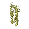
| |||||||||
| Unit cell |
| |||||||||
| Noncrystallographic symmetry (NCS) | NCS domain:
|
-Components
| #1: Protein | Mass: 18922.744 Da / Num. of mol.: 4 Source method: isolated from a genetically manipulated source Source: (gene. exp.) Thermochaetoides thermophila (fungus) / Gene: CTHT_0013690 / Production host:  #2: Chemical |   Mass: 62.068 Da / Num. of mol.: 3 / Source method: obtained synthetically / Formula: C2H6O2 Mass: 62.068 Da / Num. of mol.: 3 / Source method: obtained synthetically / Formula: C2H6O2#3: Water | ChemComp-HOH / |  Mass: 18.015 Da / Num. of mol.: 86 / Source method: isolated from a natural source / Formula: H2O Mass: 18.015 Da / Num. of mol.: 86 / Source method: isolated from a natural source / Formula: H2OHas ligand of interest | N | |
|---|
-Experimental details
-Experiment
| Experiment | Method: X-RAY DIFFRACTION / Number of used crystals: 1 |
|---|
- Sample preparation
| Crystal | Density Matthews: 3.03 Å3/Da / Density % sol: 59.46 % |
|---|---|
| Crystal grow | Temperature: 294 K / Method: vapor diffusion, sitting drop / pH: 6.5 / Details: 1.5 M Sodium Citrate pH 6.5 |
-Data collection
| Diffraction | Mean temperature: 100 K / Serial crystal experiment: N |
|---|---|
| Diffraction source | Source: SYNCHROTRON / Site: Diamond  / Beamline: I03 / Wavelength: 0.9795 Å / Beamline: I03 / Wavelength: 0.9795 Å |
| Detector | Type: DECTRIS EIGER2 XE 16M / Detector: PIXEL / Date: Nov 27, 2020 |
| Radiation | Protocol: SINGLE WAVELENGTH / Monochromatic (M) / Laue (L): M / Scattering type: x-ray |
| Radiation wavelength | Wavelength: 0.9795 Å / Relative weight: 1 |
| Reflection | Resolution: 2.4→74.78 Å / Num. obs: 35171 / % possible obs: 99.9 % / Redundancy: 6.7 % / CC1/2: 0.994 / Rmerge(I) obs: 0.091 / Rpim(I) all: 0.038 / Rrim(I) all: 0.098 / Χ2: 0.84 / Net I/σ(I): 10.6 |
| Reflection shell | Resolution: 2.4→2.49 Å / % possible obs: 99.7 % / Redundancy: 6.2 % / Rmerge(I) obs: 0.57 / Num. measured all: 22834 / Num. unique obs: 3660 / CC1/2: 0.87 / Rpim(I) all: 0.248 / Rrim(I) all: 0.623 / Χ2: 0.82 / Net I/σ(I) obs: 2.7 |
- Processing
| Software |
| ||||||||||||||||||||||||||||||||||||||||||||||||||||||||||||||||||||||||||||||||||||||||||||||||||||||||||||||||||||||||||||||||||||||||||||||||||||||||||||||||||||||||||||||||||||||
|---|---|---|---|---|---|---|---|---|---|---|---|---|---|---|---|---|---|---|---|---|---|---|---|---|---|---|---|---|---|---|---|---|---|---|---|---|---|---|---|---|---|---|---|---|---|---|---|---|---|---|---|---|---|---|---|---|---|---|---|---|---|---|---|---|---|---|---|---|---|---|---|---|---|---|---|---|---|---|---|---|---|---|---|---|---|---|---|---|---|---|---|---|---|---|---|---|---|---|---|---|---|---|---|---|---|---|---|---|---|---|---|---|---|---|---|---|---|---|---|---|---|---|---|---|---|---|---|---|---|---|---|---|---|---|---|---|---|---|---|---|---|---|---|---|---|---|---|---|---|---|---|---|---|---|---|---|---|---|---|---|---|---|---|---|---|---|---|---|---|---|---|---|---|---|---|---|---|---|---|---|---|---|---|
| Refinement | Method to determine structure: MOLECULAR REPLACEMENT / Resolution: 2.4→74.78 Å / Cor.coef. Fo:Fc: 0.936 / Cor.coef. Fo:Fc free: 0.907 / SU B: 7.842 / SU ML: 0.183 / Cross valid method: THROUGHOUT / ESU R: 0.277 / ESU R Free: 0.227 / Stereochemistry target values: MAXIMUM LIKELIHOOD / Details: HYDROGENS HAVE BEEN ADDED IN THE RIDING POSITIONS
| ||||||||||||||||||||||||||||||||||||||||||||||||||||||||||||||||||||||||||||||||||||||||||||||||||||||||||||||||||||||||||||||||||||||||||||||||||||||||||||||||||||||||||||||||||||||
| Solvent computation | Ion probe radii: 0.8 Å / Shrinkage radii: 0.8 Å / VDW probe radii: 1.2 Å / Solvent model: MASK | ||||||||||||||||||||||||||||||||||||||||||||||||||||||||||||||||||||||||||||||||||||||||||||||||||||||||||||||||||||||||||||||||||||||||||||||||||||||||||||||||||||||||||||||||||||||
| Displacement parameters | Biso mean: 51.72 Å2
| ||||||||||||||||||||||||||||||||||||||||||||||||||||||||||||||||||||||||||||||||||||||||||||||||||||||||||||||||||||||||||||||||||||||||||||||||||||||||||||||||||||||||||||||||||||||
| Refinement step | Cycle: 1 / Resolution: 2.4→74.78 Å
| ||||||||||||||||||||||||||||||||||||||||||||||||||||||||||||||||||||||||||||||||||||||||||||||||||||||||||||||||||||||||||||||||||||||||||||||||||||||||||||||||||||||||||||||||||||||
| Refine LS restraints |
|