 Movie
Movie Controller

[English] 日本語
 Yorodumi
- PDB-8onf: Crystal structure of Bdellovibrio bacteriovorus Bd2439 fibre C-te... -
+ Open data

- Basic information
| Entry | Database: PDB / ID: 8onf | ||||||
|---|---|---|---|---|---|---|---|
| Title | Crystal structure of Bdellovibrio bacteriovorus Bd2439 fibre C-terminal domains with ethylene glycol | ||||||
 Components Components | Cell wall surface anchor family protein | ||||||
 Keywords Keywords | CELL ADHESION / Adhesin / fibre / TNF / TNF-like | ||||||
| Function / homology | Cell wall surface anchor family protein Function and homology information Function and homology information | ||||||
| Biological species |  Bdellovibrio bacteriovorus HD100 (bacteria) Bdellovibrio bacteriovorus HD100 (bacteria) | ||||||
| Method |  X-RAY DIFFRACTION / SYNCHROTRON / MOLECULAR REPLACEMENT / Resolution: 1.53 Å X-RAY DIFFRACTION / SYNCHROTRON / MOLECULAR REPLACEMENT / Resolution: 1.53 Å | ||||||
 Authors Authors | Caulton, S.G. / Lovering, A.L. | ||||||
| Funding support |  United Kingdom, 1items United Kingdom, 1items
| ||||||
 Citation Citation | Journal: Nat Microbiol / Year: 2024 Title: Bdellovibrio bacteriovorus uses chimeric fibre proteins to recognize and invade a broad range of bacterial hosts. Authors: Caulton, S.G. / Lambert, C. / Tyson, J. / Radford, P. / Al-Bayati, A. / Greenwood, S. / Banks, E.J. / Clark, C. / Till, R. / Pires, E. / Sockett, R.E. / Lovering, A.L. | ||||||
| History |
|
- Structure visualization
| Structure viewer | Molecule: MolmilJmol/JSmol |
|---|
- Downloads & links
-Download
| PDBx/mmCIF format | 8onf.cif.gz | 309.7 KB | Display | PDBx/mmCIF format |
|---|---|---|---|---|
| PDB format | pdb8onf.ent.gz | 248.5 KB | Display | PDB format |
| PDBx/mmJSON format | 8onf.json.gz | Tree view | PDBx/mmJSON format | |
| Others |  Other downloads Other downloads |
-Validation report
| Arichive directory | https://data.pdbj.org/pub/pdb/validation_reports/on/8onfftp://data.pdbj.org/pub/pdb/validation_reports/on/8onf | HTTPS FTP |
|---|
-Related structure data
| Related structure data | 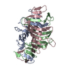 8ojnC  8ok3C  8oksC  8okwC  8ol4C 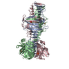 8omlC  8on4C 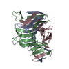 8onbC  8oncC  8ondC C: citing same article ( |
|---|---|
| Similar structure data |
-Links
 PDBj
PDBj- Assembly
| Deposited unit | 
| |||||||||
|---|---|---|---|---|---|---|---|---|---|---|
| 1 |
| |||||||||
| Unit cell |
| |||||||||
| Components on special symmetry positions |
|
-Components
| #1: Protein | Mass: 30335.496 Da / Num. of mol.: 3 Source method: isolated from a genetically manipulated source Source: (gene. exp.) Bdellovibrio bacteriovorus HD100 (bacteria)Gene: Bd2439 / Production host: #2: Chemical | ChemComp-EDO /   Mass: 62.068 Da / Num. of mol.: 7 / Source method: obtained synthetically / Formula: C2H6O2 / Feature type: SUBJECT OF INVESTIGATION Mass: 62.068 Da / Num. of mol.: 7 / Source method: obtained synthetically / Formula: C2H6O2 / Feature type: SUBJECT OF INVESTIGATION#3: Water | ChemComp-HOH / |  Mass: 18.015 Da / Num. of mol.: 937 / Source method: isolated from a natural source / Formula: H2O Mass: 18.015 Da / Num. of mol.: 937 / Source method: isolated from a natural source / Formula: H2OHas ligand of interest | Y | Has protein modification | Y | |
|---|
-Experimental details
-Experiment
| Experiment | Method: X-RAY DIFFRACTION / Number of used crystals: 1 |
|---|
- Sample preparation
| Crystal | Density Matthews: 1.95 Å3/Da / Density % sol: 36.8 % |
|---|---|
| Crystal grow | Temperature: 291.15 K / Method: vapor diffusion, sitting drop Details: 0.1 M Sodium chloride 0.1 M Sodium formate 0.1 M Bis-Tris propane 8.5 6.25 % w/v PEG 3350 6.25%w/v PEG 4000 6.25%w/v PEG 2000 6.25%w/v PEG 5000 MME |
-Data collection
| Diffraction | Mean temperature: 100 K / Serial crystal experiment: N |
|---|---|
| Diffraction source | Source: SYNCHROTRON / Site: ESRF  / Beamline: ID30B / Wavelength: 0.979 Å / Beamline: ID30B / Wavelength: 0.979 Å |
| Detector | Type: DECTRIS EIGER X 9M / Detector: PIXEL / Date: Jun 6, 2022 |
| Radiation | Protocol: SINGLE WAVELENGTH / Monochromatic (M) / Laue (L): M / Scattering type: x-ray |
| Radiation wavelength | Wavelength: 0.979 Å / Relative weight: 1 |
| Reflection | Resolution: 1.53→84.37 Å / Num. obs: 104740 / % possible obs: 96.9 % / Redundancy: 3.3 % / CC1/2: 0.998 / Rmerge(I) obs: 0.062 / Rpim(I) all: 0.037 / Rrim(I) all: 0.073 / Χ2: 0.94 / Net I/σ(I): 11.2 / Num. measured all: 341050 |
| Reflection shell | Resolution: 1.53→1.55 Å / % possible obs: 79.3 % / Redundancy: 2 % / Rmerge(I) obs: 0.653 / Num. measured all: 8323 / Num. unique obs: 4168 / CC1/2: 0.638 / Rpim(I) all: 0.526 / Rrim(I) all: 0.842 / Χ2: 0.65 / Net I/σ(I) obs: 1.1 |
- Processing
| Software |
| |||||||||||||||||||||||||||||||||||||||||||||||||||||||||||||||||||||||||||||||||||||||||||||||||||||||||||||||||||||||||||||||||||||||||||||||||||||||||||||||||||||||||||||||||||||||||||||||||||||||||||||||||||||||||||||||||
|---|---|---|---|---|---|---|---|---|---|---|---|---|---|---|---|---|---|---|---|---|---|---|---|---|---|---|---|---|---|---|---|---|---|---|---|---|---|---|---|---|---|---|---|---|---|---|---|---|---|---|---|---|---|---|---|---|---|---|---|---|---|---|---|---|---|---|---|---|---|---|---|---|---|---|---|---|---|---|---|---|---|---|---|---|---|---|---|---|---|---|---|---|---|---|---|---|---|---|---|---|---|---|---|---|---|---|---|---|---|---|---|---|---|---|---|---|---|---|---|---|---|---|---|---|---|---|---|---|---|---|---|---|---|---|---|---|---|---|---|---|---|---|---|---|---|---|---|---|---|---|---|---|---|---|---|---|---|---|---|---|---|---|---|---|---|---|---|---|---|---|---|---|---|---|---|---|---|---|---|---|---|---|---|---|---|---|---|---|---|---|---|---|---|---|---|---|---|---|---|---|---|---|---|---|---|---|---|---|---|---|---|---|---|---|---|---|---|---|---|---|---|---|---|---|---|---|
| Refinement | Method to determine structure: MOLECULAR REPLACEMENT / Resolution: 1.53→45.34 Å / SU ML: 0.17 / Cross valid method: FREE R-VALUE / σ(F): 1.34 / Phase error: 19.18 / Stereochemistry target values: ML
| |||||||||||||||||||||||||||||||||||||||||||||||||||||||||||||||||||||||||||||||||||||||||||||||||||||||||||||||||||||||||||||||||||||||||||||||||||||||||||||||||||||||||||||||||||||||||||||||||||||||||||||||||||||||||||||||||
| Solvent computation | Shrinkage radii: 0.9 Å / VDW probe radii: 1.1 Å / Solvent model: FLAT BULK SOLVENT MODEL | |||||||||||||||||||||||||||||||||||||||||||||||||||||||||||||||||||||||||||||||||||||||||||||||||||||||||||||||||||||||||||||||||||||||||||||||||||||||||||||||||||||||||||||||||||||||||||||||||||||||||||||||||||||||||||||||||
| Refinement step | Cycle: LAST / Resolution: 1.53→45.34 Å
| |||||||||||||||||||||||||||||||||||||||||||||||||||||||||||||||||||||||||||||||||||||||||||||||||||||||||||||||||||||||||||||||||||||||||||||||||||||||||||||||||||||||||||||||||||||||||||||||||||||||||||||||||||||||||||||||||
| Refine LS restraints |
| |||||||||||||||||||||||||||||||||||||||||||||||||||||||||||||||||||||||||||||||||||||||||||||||||||||||||||||||||||||||||||||||||||||||||||||||||||||||||||||||||||||||||||||||||||||||||||||||||||||||||||||||||||||||||||||||||
| LS refinement shell |
| |||||||||||||||||||||||||||||||||||||||||||||||||||||||||||||||||||||||||||||||||||||||||||||||||||||||||||||||||||||||||||||||||||||||||||||||||||||||||||||||||||||||||||||||||||||||||||||||||||||||||||||||||||||||||||||||||
| Refinement TLS params. | Method: refined / Refine-ID: X-RAY DIFFRACTION
| |||||||||||||||||||||||||||||||||||||||||||||||||||||||||||||||||||||||||||||||||||||||||||||||||||||||||||||||||||||||||||||||||||||||||||||||||||||||||||||||||||||||||||||||||||||||||||||||||||||||||||||||||||||||||||||||||
| Refinement TLS group |
|
