 Movie
Movie Controller

[English] 日本語
 Yorodumi
- PDB-8onb: Structure of the C-terminal beta helix domain of the Bdellovibrio... -
+ Open data

- Basic information
| Entry | Database: PDB / ID: 8onb | ||||||
|---|---|---|---|---|---|---|---|
| Title | Structure of the C-terminal beta helix domain of the Bdellovibrio bacteriovorus Bd3182 fibre | ||||||
 Components Components | Cell wall surface anchor family protein | ||||||
 Keywords Keywords | CELL ADHESION / Fibre / adhesin | ||||||
| Function / homology | Intramolecular chaperone auto-processing domain / Chaperone of endosialidase / Intramolecular chaperone auto-processing (ICA) domain profile. / Cell wall surface anchor family protein Function and homology information Function and homology information | ||||||
| Biological species |  Bdellovibrio bacteriovorus HD100 (bacteria) Bdellovibrio bacteriovorus HD100 (bacteria) | ||||||
| Method |  X-RAY DIFFRACTION / SYNCHROTRON / MOLECULAR REPLACEMENT / Resolution: 1.12 Å X-RAY DIFFRACTION / SYNCHROTRON / MOLECULAR REPLACEMENT / Resolution: 1.12 Å | ||||||
 Authors Authors | Caulton, S.G. / Lovering, A.L. | ||||||
| Funding support |  United Kingdom, 1items United Kingdom, 1items
| ||||||
 Citation Citation | Journal: Nat Microbiol / Year: 2024 Title: Bdellovibrio bacteriovorus uses chimeric fibre proteins to recognize and invade a broad range of bacterial hosts. Authors: Caulton, S.G. / Lambert, C. / Tyson, J. / Radford, P. / Al-Bayati, A. / Greenwood, S. / Banks, E.J. / Clark, C. / Till, R. / Pires, E. / Sockett, R.E. / Lovering, A.L. | ||||||
| History |
|
- Structure visualization
| Structure viewer | Molecule: MolmilJmol/JSmol |
|---|
- Downloads & links
-Download
| PDBx/mmCIF format | 8onb.cif.gz | 300.9 KB | Display | PDBx/mmCIF format |
|---|---|---|---|---|
| PDB format | pdb8onb.ent.gz | 246.6 KB | Display | PDB format |
| PDBx/mmJSON format | 8onb.json.gz | Tree view | PDBx/mmJSON format | |
| Others |  Other downloads Other downloads |
-Validation report
| Arichive directory | https://data.pdbj.org/pub/pdb/validation_reports/on/8onbftp://data.pdbj.org/pub/pdb/validation_reports/on/8onb | HTTPS FTP |
|---|
-Related structure data
| Related structure data | 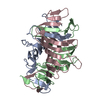 8ojnC  8ok3C  8oksC  8okwC  8ol4C 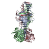 8omlC  8on4C  8oncC  8ondC  8onfC C: citing same article ( |
|---|---|
| Similar structure data |
-Links
 PDBj
PDBj- Assembly
| Deposited unit | 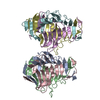
| ||||||||
|---|---|---|---|---|---|---|---|---|---|
| 1 | 
| ||||||||
| 2 | 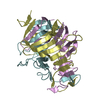
| ||||||||
| Unit cell |
|
-Components
| #1: Protein | Mass: 15519.053 Da / Num. of mol.: 6 Source method: isolated from a genetically manipulated source Source: (gene. exp.) Bdellovibrio bacteriovorus HD100 (bacteria)Gene: Bd3182 / Production host: #2: Water | ChemComp-HOH / |  Mass: 18.015 Da / Num. of mol.: 627 / Source method: isolated from a natural source / Formula: H2O Mass: 18.015 Da / Num. of mol.: 627 / Source method: isolated from a natural source / Formula: H2O |
|---|
-Experimental details
-Experiment
| Experiment | Method: X-RAY DIFFRACTION / Number of used crystals: 1 |
|---|
- Sample preparation
| Crystal | Density Matthews: 2.04 Å3/Da / Density % sol: 39.64 % |
|---|---|
| Crystal grow | Temperature: 291.15 K / Method: vapor diffusion, sitting drop Details: 0.1 M magnesium acetate tetrahydrate, 0.1 M MES pH 6.5, 10 % w/v PEG 10000 |
-Data collection
| Diffraction | Mean temperature: 100 K / Serial crystal experiment: N |
|---|---|
| Diffraction source | Source: SYNCHROTRON / Site: Diamond / Beamline: I03 / Wavelength: 0.979 Å |
| Detector | Type: DECTRIS PILATUS 6M-F / Detector: PIXEL / Date: Jan 27, 2020 |
| Radiation | Protocol: SINGLE WAVELENGTH / Monochromatic (M) / Laue (L): M / Scattering type: x-ray |
| Radiation wavelength | Wavelength: 0.979 Å / Relative weight: 1 |
| Reflection | Resolution: 1.12→84.35 Å / Num. obs: 176672 / % possible obs: 61.3 % / Redundancy: 6.8 % / CC1/2: 0.999 / Net I/σ(I): 16.4 |
| Reflection shell | Resolution: 1.12→1.241 Å / Num. unique obs: 8836 / CC1/2: 0.706 |
- Processing
| Software |
| |||||||||||||||||||||||||||||||||||||||||||||||||||||||||||||||||||||||||||||||||||||||||||||||||||||||||||||||||||||||||||||||||||||||||||||||||||||||||||||||||||||||||||||||||||||||||||||||||||||||||||||||||||||||||
|---|---|---|---|---|---|---|---|---|---|---|---|---|---|---|---|---|---|---|---|---|---|---|---|---|---|---|---|---|---|---|---|---|---|---|---|---|---|---|---|---|---|---|---|---|---|---|---|---|---|---|---|---|---|---|---|---|---|---|---|---|---|---|---|---|---|---|---|---|---|---|---|---|---|---|---|---|---|---|---|---|---|---|---|---|---|---|---|---|---|---|---|---|---|---|---|---|---|---|---|---|---|---|---|---|---|---|---|---|---|---|---|---|---|---|---|---|---|---|---|---|---|---|---|---|---|---|---|---|---|---|---|---|---|---|---|---|---|---|---|---|---|---|---|---|---|---|---|---|---|---|---|---|---|---|---|---|---|---|---|---|---|---|---|---|---|---|---|---|---|---|---|---|---|---|---|---|---|---|---|---|---|---|---|---|---|---|---|---|---|---|---|---|---|---|---|---|---|---|---|---|---|---|---|---|---|---|---|---|---|---|---|---|---|---|---|---|---|---|
| Refinement | Method to determine structure: MOLECULAR REPLACEMENT / Resolution: 1.12→84.35 Å / SU ML: 0.1 / Cross valid method: FREE R-VALUE / σ(F): 1.35 / Phase error: 23.49 / Stereochemistry target values: ML
| |||||||||||||||||||||||||||||||||||||||||||||||||||||||||||||||||||||||||||||||||||||||||||||||||||||||||||||||||||||||||||||||||||||||||||||||||||||||||||||||||||||||||||||||||||||||||||||||||||||||||||||||||||||||||
| Solvent computation | Shrinkage radii: 0.9 Å / VDW probe radii: 1.1 Å / Solvent model: FLAT BULK SOLVENT MODEL | |||||||||||||||||||||||||||||||||||||||||||||||||||||||||||||||||||||||||||||||||||||||||||||||||||||||||||||||||||||||||||||||||||||||||||||||||||||||||||||||||||||||||||||||||||||||||||||||||||||||||||||||||||||||||
| Refinement step | Cycle: LAST / Resolution: 1.12→84.35 Å
| |||||||||||||||||||||||||||||||||||||||||||||||||||||||||||||||||||||||||||||||||||||||||||||||||||||||||||||||||||||||||||||||||||||||||||||||||||||||||||||||||||||||||||||||||||||||||||||||||||||||||||||||||||||||||
| Refine LS restraints |
| |||||||||||||||||||||||||||||||||||||||||||||||||||||||||||||||||||||||||||||||||||||||||||||||||||||||||||||||||||||||||||||||||||||||||||||||||||||||||||||||||||||||||||||||||||||||||||||||||||||||||||||||||||||||||
| LS refinement shell |
|
