 Movie
Movie Controller

[English] 日本語
 Yorodumi
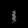
- PDB-8oi8: Cryo-EM structure of ADP-bound, filamentous beta-actin harboring ... -
+ Open data

- Basic information
| Entry | Database: PDB / ID: 8oi8 | ||||||||||||
|---|---|---|---|---|---|---|---|---|---|---|---|---|---|
| Title | Cryo-EM structure of ADP-bound, filamentous beta-actin harboring the R183W mutation | ||||||||||||
 Components Components | Actin, cytoplasmic 1 | ||||||||||||
 Keywords Keywords | STRUCTURAL PROTEIN / Actin filament / cytoskeletal protein / ATPase | ||||||||||||
| Function / homology |  Function and homology information Function and homology informationpositive regulation of norepinephrine uptake / bBAF complex / cellular response to cytochalasin B / Formation of the embryonic stem cell BAF (esBAF) complex / npBAF complex / brahma complex / nBAF complex / Formation of the canonical BAF (cBAF) complex / regulation of transepithelial transport / morphogenesis of a polarized epithelium ...positive regulation of norepinephrine uptake / bBAF complex / cellular response to cytochalasin B / Formation of the embryonic stem cell BAF (esBAF) complex / npBAF complex / brahma complex / nBAF complex / Formation of the canonical BAF (cBAF) complex / regulation of transepithelial transport / morphogenesis of a polarized epithelium / Formation of the polybromo-BAF (pBAF) complex / Formation of neuronal progenitor and neuronal BAF (npBAF and nBAF) / structural constituent of postsynaptic actin cytoskeleton / Formation of annular gap junctions / Formation of the dystrophin-glycoprotein complex (DGC) / GBAF complex / Gap junction degradation / Formation of the non-canonical BAF (ncBAF) complex / protein localization to adherens junction / regulation of G0 to G1 transition / Cell-extracellular matrix interactions / dense body / Folding of actin by CCT/TriC / Tat protein binding / postsynaptic actin cytoskeleton / RSC-type complex / Regulation of CDH1 Function / regulation of double-strand break repair / Prefoldin mediated transfer of substrate to CCT/TriC / regulation of nucleotide-excision repair / Adherens junctions interactions / adherens junction assembly / RHOF GTPase cycle / apical protein localization / Sensory processing of sound by outer hair cells of the cochlea / regulation of mitotic metaphase/anaphase transition / tight junction / SWI/SNF complex / Sensory processing of sound by inner hair cells of the cochlea / Interaction between L1 and Ankyrins / positive regulation of T cell differentiation / apical junction complex / positive regulation of double-strand break repair / maintenance of blood-brain barrier / regulation of norepinephrine uptake / transporter regulator activity / positive regulation of stem cell population maintenance / NuA4 histone acetyltransferase complex / Recycling pathway of L1 / Regulation of MITF-M-dependent genes involved in pigmentation / cortical cytoskeleton / establishment or maintenance of cell polarity / nitric-oxide synthase binding / regulation of G1/S transition of mitotic cell cycle / brush border / EPH-ephrin mediated repulsion of cells / negative regulation of cell differentiation / regulation of synaptic vesicle endocytosis / positive regulation of myoblast differentiation / RHO GTPases Activate WASPs and WAVEs / kinesin binding / regulation of protein localization to plasma membrane / RHO GTPases activate IQGAPs / positive regulation of double-strand break repair via homologous recombination / EPHB-mediated forward signaling / cytoskeleton organization / axonogenesis / substantia nigra development / calyx of Held / nitric-oxide synthase regulator activity / FCGR3A-mediated phagocytosis / Translocation of SLC2A4 (GLUT4) to the plasma membrane / actin filament / adherens junction / positive regulation of cell differentiation / cell motility / Regulation of endogenous retroelements by Piwi-interacting RNAs (piRNAs) / RHO GTPases Activate Formins / Signaling by high-kinase activity BRAF mutants / MAP2K and MAPK activation / Regulation of actin dynamics for phagocytic cup formation / structural constituent of cytoskeleton / kinetochore / VEGFA-VEGFR2 Pathway / B-WICH complex positively regulates rRNA expression / platelet aggregation / tau protein binding / Hydrolases; Acting on acid anhydrides; Acting on acid anhydrides to facilitate cellular and subcellular movement / DNA Damage Recognition in GG-NER / Schaffer collateral - CA1 synapse / nuclear matrix / Signaling by RAF1 mutants / cell-cell junction / Signaling by moderate kinase activity BRAF mutants / Paradoxical activation of RAF signaling by kinase inactive BRAF / Signaling downstream of RAS mutants / cytoplasmic ribonucleoprotein granule / Signaling by BRAF and RAF1 fusions / UCH proteinases / nucleosome Similarity search - Function | ||||||||||||
| Biological species |  Homo sapiens (human) Homo sapiens (human) | ||||||||||||
| Method | ELECTRON MICROSCOPY / single particle reconstruction / cryo EM / Resolution: 2.28 Å | ||||||||||||
 Authors Authors | Oosterheert, W. / Blanc, F.E.C. / Roy, A. / Belyy, A. / Hofnagel, O. / Hummer, G. / Bieling, P. / Raunser, S. | ||||||||||||
| Funding support |  Germany, European Union, 3items Germany, European Union, 3items
| ||||||||||||
 Citation Citation | Journal: Nat Struct Mol Biol / Year: 2023 Title: Molecular mechanisms of inorganic-phosphate release from the core and barbed end of actin filaments. Authors: Wout Oosterheert / Florian E C Blanc / Ankit Roy / Alexander Belyy / Micaela Boiero Sanders / Oliver Hofnagel / Gerhard Hummer / Peter Bieling / Stefan Raunser / Abstract: The release of inorganic phosphate (P) from actin filaments constitutes a key step in their regulated turnover, which is fundamental to many cellular functions. The mechanisms underlying P release ...The release of inorganic phosphate (P) from actin filaments constitutes a key step in their regulated turnover, which is fundamental to many cellular functions. The mechanisms underlying P release from the core and barbed end of actin filaments remain unclear. Here, using human and bovine actin isoforms, we combine cryo-EM with molecular-dynamics simulations and in vitro reconstitution to demonstrate how actin releases P through a 'molecular backdoor'. While constantly open at the barbed end, the backdoor is predominantly closed in filament-core subunits and opens only transiently through concerted amino acid rearrangements. This explains why P escapes rapidly from the filament end but slowly from internal subunits. In a nemaline-myopathy-associated actin variant, the backdoor is predominantly open in filament-core subunits, resulting in accelerated P release and filaments with drastically shortened ADP-P caps. Our results provide the molecular basis for P release from actin and exemplify how a disease-linked mutation distorts the nucleotide-state distribution and atomic structure of the filament. | ||||||||||||
| History |
|
- Structure visualization
| Structure viewer | Molecule: MolmilJmol/JSmol |
|---|
- Downloads & links
-Download
| PDBx/mmCIF format | 8oi8.cif.gz | 397.5 KB | Display | PDBx/mmCIF format |
|---|---|---|---|---|
| PDB format | pdb8oi8.ent.gz | 271.4 KB | Display | PDB format |
| PDBx/mmJSON format | 8oi8.json.gz | Tree view | PDBx/mmJSON format | |
| Others |  Other downloads Other downloads |
-Validation report
| Arichive directory | https://data.pdbj.org/pub/pdb/validation_reports/oi/8oi8ftp://data.pdbj.org/pub/pdb/validation_reports/oi/8oi8 | HTTPS FTP |
|---|
-Related structure data
| Related structure data | 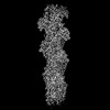 16888MC 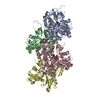 8oi6C 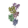 8oidC C: citing same article ( M: map data used to model this data |
|---|---|
| Similar structure data |
-Links
 PDBj
PDBj


- Assembly
| Deposited unit | 
| ||||||||||||||||||||||||||||||||||||||||||||||||||||||||||||||||||||||||||||||||||||||||||||||||||||||||||||||||||||||||||||||||||||||||||||||||||||
|---|---|---|---|---|---|---|---|---|---|---|---|---|---|---|---|---|---|---|---|---|---|---|---|---|---|---|---|---|---|---|---|---|---|---|---|---|---|---|---|---|---|---|---|---|---|---|---|---|---|---|---|---|---|---|---|---|---|---|---|---|---|---|---|---|---|---|---|---|---|---|---|---|---|---|---|---|---|---|---|---|---|---|---|---|---|---|---|---|---|---|---|---|---|---|---|---|---|---|---|---|---|---|---|---|---|---|---|---|---|---|---|---|---|---|---|---|---|---|---|---|---|---|---|---|---|---|---|---|---|---|---|---|---|---|---|---|---|---|---|---|---|---|---|---|---|---|---|---|---|
| 1 |
| ||||||||||||||||||||||||||||||||||||||||||||||||||||||||||||||||||||||||||||||||||||||||||||||||||||||||||||||||||||||||||||||||||||||||||||||||||||
| Noncrystallographic symmetry (NCS) | NCS domain:
NCS domain segments: Ens-ID: ens_1
NCS oper:
|
-Components
| #1: Protein | Mass: 41792.633 Da / Num. of mol.: 5 / Mutation: R183W, C272A Source method: isolated from a genetically manipulated source Source: (gene. exp.) Homo sapiens (human) / Gene: ACTB / Plasmid: p2920 pFL_ACTB_R183W_C272A / Cell line (production host): BTI-Tnao38 / Production host:  Trichoplusia ni (cabbage looper) Trichoplusia ni (cabbage looper)References: UniProt: P60709, Hydrolases; Acting on acid anhydrides; Acting on acid anhydrides to facilitate cellular and subcellular movement #2: Chemical | ChemComp-ADP /   Mass: 427.201 Da / Num. of mol.: 5 / Source method: obtained synthetically / Formula: C10H15N5O10P2 / Feature type: SUBJECT OF INVESTIGATION / Comment: ADP, energy-carrying molecule*YM Mass: 427.201 Da / Num. of mol.: 5 / Source method: obtained synthetically / Formula: C10H15N5O10P2 / Feature type: SUBJECT OF INVESTIGATION / Comment: ADP, energy-carrying molecule*YM#3: Chemical | ChemComp-MG /   Mass: 24.305 Da / Num. of mol.: 5 / Source method: obtained synthetically / Formula: Mg / Feature type: SUBJECT OF INVESTIGATION Mass: 24.305 Da / Num. of mol.: 5 / Source method: obtained synthetically / Formula: Mg / Feature type: SUBJECT OF INVESTIGATION#4: Water | ChemComp-HOH / |  Mass: 18.015 Da / Num. of mol.: 585 / Source method: isolated from a natural source / Formula: H2O Mass: 18.015 Da / Num. of mol.: 585 / Source method: isolated from a natural source / Formula: H2OHas ligand of interest | Y | |
|---|
-Experimental details
-Experiment
| Experiment | Method: ELECTRON MICROSCOPY |
|---|---|
| EM experiment | Aggregation state: FILAMENT / 3D reconstruction method: single particle reconstruction |
- Sample preparation
| Component | Name: Actin filament harboring the R183W mutation. / Type: COMPLEX Details: Actin was purified as monomer from insect cells. It was then polymerized into a filament in vitro. Entity ID: #1 / Source: RECOMBINANT | ||||||||||||||||||||||||||||||
|---|---|---|---|---|---|---|---|---|---|---|---|---|---|---|---|---|---|---|---|---|---|---|---|---|---|---|---|---|---|---|---|
| Molecular weight | Experimental value: NO | ||||||||||||||||||||||||||||||
| Source (natural) | Organism: Homo sapiens (human) | ||||||||||||||||||||||||||||||
| Source (recombinant) | Organism: Trichoplusia ni (cabbage looper) / Cell: BTI-Tnao38 | ||||||||||||||||||||||||||||||
| Buffer solution | pH: 7.1 Details: 1x KMEH (10 mM HEPES pH 7.1, 100 mM KCl, 2 mM MgCl2, 1 mM EGTA) supplemented with 0.02% Tween20 (v/v) | ||||||||||||||||||||||||||||||
| Buffer component |
| ||||||||||||||||||||||||||||||
| Specimen | Conc.: 2.3 mg/ml / Embedding applied: NO / Shadowing applied: NO / Staining applied: NO / Vitrification applied: YES Details: Actin filaments were reconstituted by adding salt to monomeric actin. | ||||||||||||||||||||||||||||||
| Specimen support | Grid material: COPPER / Grid mesh size: 300 divisions/in. / Grid type: Quantifoil R2/1 | ||||||||||||||||||||||||||||||
| Vitrification | Instrument: FEI VITROBOT MARK IV / Cryogen name: ETHANE / Humidity: 100 % / Chamber temperature: 286 K |
- Electron microscopy imaging
| Experimental equipment |  Model: Titan Krios / Image courtesy: FEI Company |
|---|---|
| Microscopy | Model: FEI TITAN KRIOS Details: Titan Krios G3 microscope was aligned using Sherpa (FEI). Data collected in superresolution mode. |
| Electron gun | Electron source:  FIELD EMISSION GUN / Accelerating voltage: 300 kV / Illumination mode: SPOT SCAN FIELD EMISSION GUN / Accelerating voltage: 300 kV / Illumination mode: SPOT SCAN |
| Electron lens | Mode: BRIGHT FIELD / Nominal magnification: 130000 X / Nominal defocus max: 2000 nm / Nominal defocus min: 800 nm / Cs: 2.7 mm / C2 aperture diameter: 50 µm / Alignment procedure: ZEMLIN TABLEAU |
| Specimen holder | Cryogen: NITROGEN / Specimen holder model: FEI TITAN KRIOS AUTOGRID HOLDER |
| Image recording | Electron dose: 70 e/Å2 / Film or detector model: GATAN K3 BIOQUANTUM (6k x 4k) / Num. of grids imaged: 1 / Num. of real images: 7916 |
| EM imaging optics | Energyfilter name: GIF Bioquantum / Energyfilter slit width: 15 eV |
- Processing
| Software |
| ||||||||||||||||||||||||||||||||||||||||
|---|---|---|---|---|---|---|---|---|---|---|---|---|---|---|---|---|---|---|---|---|---|---|---|---|---|---|---|---|---|---|---|---|---|---|---|---|---|---|---|---|---|
| EM software |
| ||||||||||||||||||||||||||||||||||||||||
| Image processing | Details: K3 operated in super-resolution mode. | ||||||||||||||||||||||||||||||||||||||||
| CTF correction | Type: PHASE FLIPPING AND AMPLITUDE CORRECTION | ||||||||||||||||||||||||||||||||||||||||
| Particle selection | Num. of particles selected: 1569882 | ||||||||||||||||||||||||||||||||||||||||
| Symmetry | Point symmetry: C1 (asymmetric) | ||||||||||||||||||||||||||||||||||||||||
| 3D reconstruction | Resolution: 2.28 Å / Resolution method: FSC 0.143 CUT-OFF / Num. of particles: 1386604 / Details: Refinement from Local searches in RELION. / Symmetry type: POINT | ||||||||||||||||||||||||||||||||||||||||
| Atomic model building | Protocol: FLEXIBLE FIT / Space: REAL Details: The structure was refined using phenix-real-space refine. | ||||||||||||||||||||||||||||||||||||||||
| Atomic model building | Accession code: 8a2t / Source name: PDB / Type: experimental model | ||||||||||||||||||||||||||||||||||||||||
| Refinement | Cross valid method: NONE Stereochemistry target values: GeoStd + Monomer Library + CDL v1.2 | ||||||||||||||||||||||||||||||||||||||||
| Displacement parameters | Biso mean: 31.83 Å2 | ||||||||||||||||||||||||||||||||||||||||
| Refine LS restraints |
| ||||||||||||||||||||||||||||||||||||||||
| Refine LS restraints NCS |
|
