Movie
Movie Controller

[English] 日本語
 Yorodumi
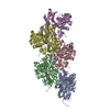
- EMDB-16889: Cryo-EM structure of ADP-bound, filamentous beta-actin harboring ... -
+ Open data

- Basic information
| Entry | 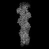 | ||||||||||||
|---|---|---|---|---|---|---|---|---|---|---|---|---|---|
| Title | Cryo-EM structure of ADP-bound, filamentous beta-actin harboring the N111S mutation | ||||||||||||
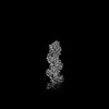
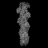
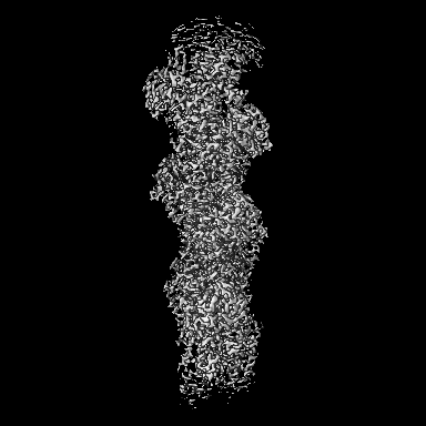
 Map data Map data | Sharpened, local-resolution filtered cryo-EM density map of filamentous beta-actin harboring the N111S mutation. | ||||||||||||
 Sample Sample |
| ||||||||||||
 Keywords Keywords | Actin filament / cytoskeletal protein / ATPase / STRUCTURAL PROTEIN | ||||||||||||
| Function / homology |  Function and homology information Function and homology informationpositive regulation of norepinephrine uptake / bBAF complex / cellular response to cytochalasin B / Formation of the embryonic stem cell BAF (esBAF) complex / npBAF complex / brahma complex / nBAF complex / Formation of the canonical BAF (cBAF) complex / regulation of transepithelial transport / morphogenesis of a polarized epithelium ...positive regulation of norepinephrine uptake / bBAF complex / cellular response to cytochalasin B / Formation of the embryonic stem cell BAF (esBAF) complex / npBAF complex / brahma complex / nBAF complex / Formation of the canonical BAF (cBAF) complex / regulation of transepithelial transport / morphogenesis of a polarized epithelium / Formation of the polybromo-BAF (pBAF) complex / Formation of neuronal progenitor and neuronal BAF (npBAF and nBAF) / structural constituent of postsynaptic actin cytoskeleton / Formation of annular gap junctions / Formation of the dystrophin-glycoprotein complex (DGC) / GBAF complex / Gap junction degradation / Formation of the non-canonical BAF (ncBAF) complex / protein localization to adherens junction / regulation of G0 to G1 transition / Cell-extracellular matrix interactions / dense body / Folding of actin by CCT/TriC / Tat protein binding / postsynaptic actin cytoskeleton / RSC-type complex / Regulation of CDH1 Function / regulation of double-strand break repair / Prefoldin mediated transfer of substrate to CCT/TriC / regulation of nucleotide-excision repair / Adherens junctions interactions / adherens junction assembly / RHOF GTPase cycle / apical protein localization / Sensory processing of sound by outer hair cells of the cochlea / tight junction / regulation of mitotic metaphase/anaphase transition / SWI/SNF complex / Sensory processing of sound by inner hair cells of the cochlea / positive regulation of T cell differentiation / Interaction between L1 and Ankyrins / apical junction complex / maintenance of blood-brain barrier / positive regulation of stem cell population maintenance / regulation of norepinephrine uptake / transporter regulator activity / NuA4 histone acetyltransferase complex / positive regulation of double-strand break repair / Recycling pathway of L1 / Regulation of MITF-M-dependent genes involved in pigmentation / cortical cytoskeleton / establishment or maintenance of cell polarity / nitric-oxide synthase binding / brush border / EPH-ephrin mediated repulsion of cells / negative regulation of cell differentiation / regulation of synaptic vesicle endocytosis / positive regulation of myoblast differentiation / RHO GTPases Activate WASPs and WAVEs / kinesin binding / regulation of protein localization to plasma membrane / RHO GTPases activate IQGAPs / positive regulation of double-strand break repair via homologous recombination / regulation of G1/S transition of mitotic cell cycle / axonogenesis / cytoskeleton organization / EPHB-mediated forward signaling / substantia nigra development / calyx of Held / nitric-oxide synthase regulator activity / cell motility / FCGR3A-mediated phagocytosis / Translocation of SLC2A4 (GLUT4) to the plasma membrane / actin filament / adherens junction / positive regulation of cell differentiation / Regulation of endogenous retroelements by Piwi-interacting RNAs (piRNAs) / RHO GTPases Activate Formins / Signaling by high-kinase activity BRAF mutants / MAP2K and MAPK activation / Regulation of actin dynamics for phagocytic cup formation / VEGFA-VEGFR2 Pathway / B-WICH complex positively regulates rRNA expression / structural constituent of cytoskeleton / platelet aggregation / kinetochore / tau protein binding / Hydrolases; Acting on acid anhydrides; Acting on acid anhydrides to facilitate cellular and subcellular movement / DNA Damage Recognition in GG-NER / Schaffer collateral - CA1 synapse / nuclear matrix / cytoplasmic ribonucleoprotein granule / Signaling by RAF1 mutants / cell-cell junction / Signaling by moderate kinase activity BRAF mutants / Paradoxical activation of RAF signaling by kinase inactive BRAF / Signaling downstream of RAS mutants / Signaling by BRAF and RAF1 fusions / UCH proteinases / nucleosome Similarity search - Function | ||||||||||||
| Biological species |  Homo sapiens (human) Homo sapiens (human) | ||||||||||||
| Method | single particle reconstruction / cryo EM / Resolution: 2.3 Å | ||||||||||||
 Authors Authors | Oosterheert W / Blanc FEC / Roy A / Belyy A / Hofnagel O / Hummer G / Bieling P / Raunser S | ||||||||||||
| Funding support |  Germany, European Union, 3 items Germany, European Union, 3 items
| ||||||||||||
 Citation Citation | Journal: Nat Struct Mol Biol / Year: 2023 Title: Molecular mechanisms of inorganic-phosphate release from the core and barbed end of actin filaments. Authors: Wout Oosterheert / Florian E C Blanc / Ankit Roy / Alexander Belyy / Micaela Boiero Sanders / Oliver Hofnagel / Gerhard Hummer / Peter Bieling / Stefan Raunser / Abstract: The release of inorganic phosphate (P) from actin filaments constitutes a key step in their regulated turnover, which is fundamental to many cellular functions. The mechanisms underlying P release ...The release of inorganic phosphate (P) from actin filaments constitutes a key step in their regulated turnover, which is fundamental to many cellular functions. The mechanisms underlying P release from the core and barbed end of actin filaments remain unclear. Here, using human and bovine actin isoforms, we combine cryo-EM with molecular-dynamics simulations and in vitro reconstitution to demonstrate how actin releases P through a 'molecular backdoor'. While constantly open at the barbed end, the backdoor is predominantly closed in filament-core subunits and opens only transiently through concerted amino acid rearrangements. This explains why P escapes rapidly from the filament end but slowly from internal subunits. In a nemaline-myopathy-associated actin variant, the backdoor is predominantly open in filament-core subunits, resulting in accelerated P release and filaments with drastically shortened ADP-P caps. Our results provide the molecular basis for P release from actin and exemplify how a disease-linked mutation distorts the nucleotide-state distribution and atomic structure of the filament. | ||||||||||||
| History |
|
- Structure visualization
| Supplemental images |
|---|

- Downloads & links
-EMDB archive
| Map data | emd_16889.map.gz | 129.5 MB | EMDB map data format | |
|---|---|---|---|---|
| Header (meta data) | emd-16889-v30.xmlemd-16889.xml | 25.6 KB 25.6 KB | Display Display | EMDB header |
| FSC (resolution estimation) | emd_16889_fsc.xml | 13.6 KB | Display | FSC data file |
| Images | 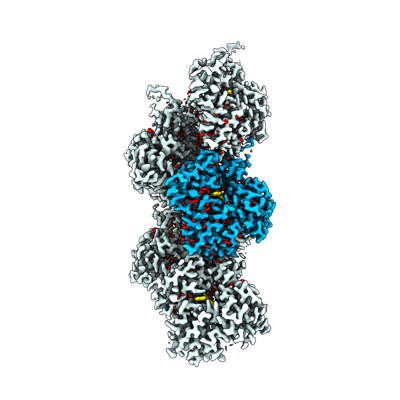 emd_16889.png emd_16889.png | 89.7 KB | ||
| Masks | emd_16889_msk_1.map | 216 MB | Mask map | |
| Filedesc metadata | emd-16889.cif.gz | 7.8 KB | ||
| Others | emd_16889_additional_1.map.gzemd_16889_half_map_1.map.gzemd_16889_half_map_2.map.gz | 168.6 MB 170.5 MB 170.5 MB | ||
| Archive directory |  http://ftp.pdbj.org/pub/emdb/structures/EMD-16889ftp://ftp.pdbj.org/pub/emdb/structures/EMD-16889 http://ftp.pdbj.org/pub/emdb/structures/EMD-16889ftp://ftp.pdbj.org/pub/emdb/structures/EMD-16889 | HTTPS FTP |
-Related structure data
| Related structure data |  8oidMC 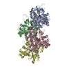 8oi6C  8oi8C C: citing same article ( M: atomic model generated by this map |
|---|---|
| Similar structure data |
-Links
| EMDB pages | EMDB (EBI/PDBe) / EMDataResource |
|---|---|
| Related items in Molecule of the Month |


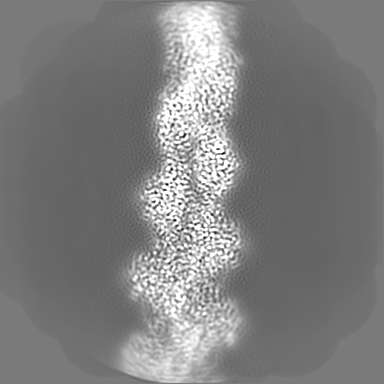
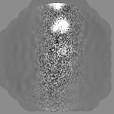
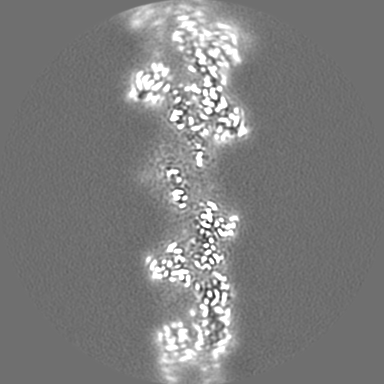
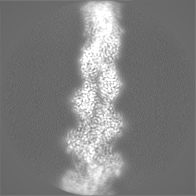
-Map
| File | Download / File: emd_16889.map.gz / Format: CCP4 / Size: 216 MB / Type: IMAGE STORED AS FLOATING POINT NUMBER (4 BYTES) | ||||||||||||||||||||||||||||||||||||
|---|---|---|---|---|---|---|---|---|---|---|---|---|---|---|---|---|---|---|---|---|---|---|---|---|---|---|---|---|---|---|---|---|---|---|---|---|---|
| Annotation | Sharpened, local-resolution filtered cryo-EM density map of filamentous beta-actin harboring the N111S mutation. | ||||||||||||||||||||||||||||||||||||
| Projections & slices | Image control
Images are generated by Spider. | ||||||||||||||||||||||||||||||||||||
| Voxel size | X=Y=Z: 0.695 Å | ||||||||||||||||||||||||||||||||||||
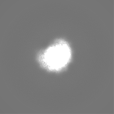
| Density |
| ||||||||||||||||||||||||||||||||||||
| Symmetry | Space group: 1 | ||||||||||||||||||||||||||||||||||||
| Details | EMDB XML:
|
 Z (Sec.)
 Y (Row.)
 X (Col.)


-Supplemental data
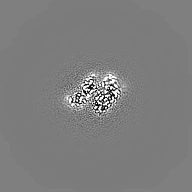
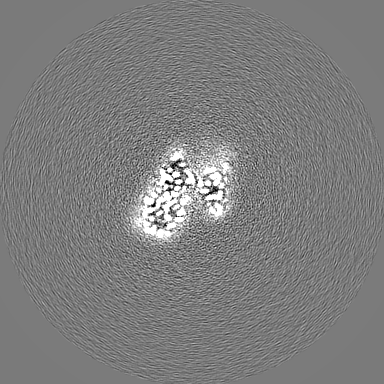
-Mask #1
| File | emd_16889_msk_1.map | ||||||||||||
|---|---|---|---|---|---|---|---|---|---|---|---|---|---|
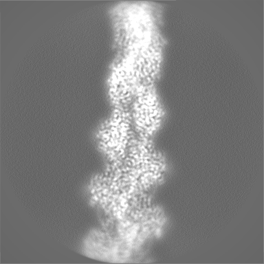
| Projections & Slices |
| ||||||||||||
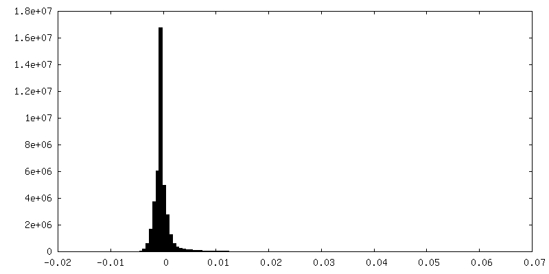
| Density Histograms |


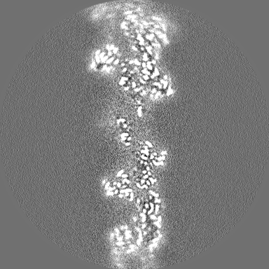
-Additional map: 3D refined, unsharpened cryo-EM density map of filamentous...
| File | emd_16889_additional_1.map | ||||||||||||
|---|---|---|---|---|---|---|---|---|---|---|---|---|---|
| Annotation | 3D refined, unsharpened cryo-EM density map of filamentous beta-actin harboring the N111S mutation. | ||||||||||||
| Projections & Slices |
| ||||||||||||
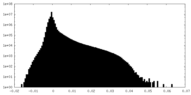
| Density Histograms |


-Half map: Half map 1 of the refinement of filamentous...
| File | emd_16889_half_map_1.map | ||||||||||||
|---|---|---|---|---|---|---|---|---|---|---|---|---|---|
| Annotation | Half map 1 of the refinement of filamentous beta-actin harboring the N111S mutation. | ||||||||||||
| Projections & Slices |
| ||||||||||||
| Density Histograms |


-Half map: Half map 2 of the refinement of filamentous...
| File | emd_16889_half_map_2.map | ||||||||||||
|---|---|---|---|---|---|---|---|---|---|---|---|---|---|
| Annotation | Half map 2 of the refinement of filamentous beta-actin harboring the N111S mutation. | ||||||||||||
| Projections & Slices |
| ||||||||||||
| Density Histograms |


- Sample components
-Entire : Actin filament harboring the N111S mutation.
| Entire | Name: Actin filament harboring the N111S mutation. |
|---|---|
| Components |
|
-Supramolecule #1: Actin filament harboring the N111S mutation.
| Supramolecule | Name: Actin filament harboring the N111S mutation. / type: complex / ID: 1 / Parent: 0 / Macromolecule list: #1 Details: Beta-actin was expressed as fusion protein, with thymosin beta4 and a deca-His-tag fused to the actin C-terminus. During the purification, thymosin beta-4 and the deca-His-tag were removed. ...Details: Beta-actin was expressed as fusion protein, with thymosin beta4 and a deca-His-tag fused to the actin C-terminus. During the purification, thymosin beta-4 and the deca-His-tag were removed. Actin was purified as monomer from insect cells. It was then polymerized into a filament in vitro. |
|---|---|
| Source (natural) | Organism: Homo sapiens (human) |
-Macromolecule #1: Actin, cytoplasmic 1, N-terminally processed
| Macromolecule | Name: Actin, cytoplasmic 1, N-terminally processed / type: protein_or_peptide / ID: 1 / Number of copies: 5 / Enantiomer: LEVO |
|---|---|
| Source (natural) | Organism: Homo sapiens (human) |
| Molecular weight | Theoretical: 41.73659 KDa |
| Recombinant expression | Organism:  Trichoplusia ni (cabbage looper) Trichoplusia ni (cabbage looper) |
| Sequence | String: MDDDIAALVV DNGSGMCKAG FAGDDAPRAV FPSIVGRPRH QGVMVGMGQK DSYVGDEAQS KRGILTLKYP IE(HIC)GIV TNW DDMEKIWHHT FYNELRVAPE EHPVLLTEAP LSPKANREKM TQIMFETFNT PAMYVAIQAV LSLYASGRTT GIVMDSG DG VTHTVPIYEG ...String: MDDDIAALVV DNGSGMCKAG FAGDDAPRAV FPSIVGRPRH QGVMVGMGQK DSYVGDEAQS KRGILTLKYP IE(HIC)GIV TNW DDMEKIWHHT FYNELRVAPE EHPVLLTEAP LSPKANREKM TQIMFETFNT PAMYVAIQAV LSLYASGRTT GIVMDSG DG VTHTVPIYEG YALPHAILRL DLAGRDLTDY LMKILTERGY SFTTTAEREI VRDIKEKLCY VALDFEQEMA TAASSSSL E KSYELPDGQV ITIGNERFRC PEALFQPSFL GMESAGIHET TFNSIMKCDV DIRKDLYANT VLSGGTTMYP GIADRMQKE ITALAPSTMK IKIIAPPERK YSVWIGGSIL ASLSTFQQMW ISKQEYDESG PSIVHRKCF UniProtKB: Actin, cytoplasmic 1 |
-Macromolecule #2: ADENOSINE-5'-DIPHOSPHATE
| Macromolecule | Name: ADENOSINE-5'-DIPHOSPHATE / type: ligand / ID: 2 / Number of copies: 5 / Formula: ADP |
|---|---|
| Molecular weight | Theoretical: 427.201 Da |
| Chemical component information |  ChemComp-ADP: |
-Macromolecule #3: MAGNESIUM ION
| Macromolecule | Name: MAGNESIUM ION / type: ligand / ID: 3 / Number of copies: 5 / Formula: MG |
|---|---|
| Molecular weight | Theoretical: 24.305 Da |
-Macromolecule #4: water
| Macromolecule | Name: water / type: ligand / ID: 4 / Number of copies: 616 / Formula: HOH |
|---|---|
| Molecular weight | Theoretical: 18.015 Da |
| Chemical component information |  ChemComp-HOH: |
-Experimental details
-Structure determination
| Method | cryo EM |
|---|---|
 Processing Processing | single particle reconstruction |
| Aggregation state | filament |
-Sample preparation
| Concentration | 2.5 mg/mL | ||||||||||||||||||
|---|---|---|---|---|---|---|---|---|---|---|---|---|---|---|---|---|---|---|---|
| Buffer | pH: 7.1 Component:
Details: 1x KMEH (10 mM HEPES pH 7.1, 100 mM KCl, 2 mM MgCl2, 1 mM EGTA) supplemented with 0.02% Tween20 (v/v) | ||||||||||||||||||
| Grid | Model: Quantifoil R2/1 / Material: COPPER / Mesh: 300 / Support film - Material: CARBON / Support film - topology: HOLEY / Pretreatment - Type: GLOW DISCHARGE / Pretreatment - Time: 90 sec. | ||||||||||||||||||
| Vitrification | Cryogen name: ETHANE-PROPANE / Chamber humidity: 100 % / Chamber temperature: 286 K / Instrument: FEI VITROBOT MARK IV | ||||||||||||||||||
| Details | Actin filaments were reconstituted by adding salt to monomeric actin. |
- Electron microscopy
| Microscope | FEI TITAN KRIOS |
|---|---|
| Specialist optics | Energy filter - Name: GIF Bioquantum / Energy filter - Slit width: 15 eV |
| Details | Titan Krios G3 microscope was aligned using Sherpa (FEI). Data collected in superresolution mode. |
| Image recording | Film or detector model: GATAN K3 BIOQUANTUM (6k x 4k) / Number grids imaged: 1 / Number real images: 9516 / Average electron dose: 70.0 e/Å2 |
| Electron beam | Acceleration voltage: 300 kV / Electron source:  FIELD EMISSION GUN FIELD EMISSION GUN |
| Electron optics | C2 aperture diameter: 50.0 µm / Illumination mode: SPOT SCAN / Imaging mode: BRIGHT FIELD / Cs: 2.7 mm / Nominal defocus max: 2.0 µm / Nominal defocus min: 0.8 µm / Nominal magnification: 130000 |
| Sample stage | Specimen holder model: FEI TITAN KRIOS AUTOGRID HOLDER / Cooling holder cryogen: NITROGEN |
| Experimental equipment |  Model: Titan Krios / Image courtesy: FEI Company |
+Image processing


-Atomic model buiding 1
| Initial model | PDB ID: Chain - Source name: PDB / Chain - Initial model type: experimental model |
|---|---|
| Details | chain C of pdb 8A2T (including all water molecules) was fit in the central actin subunit of the density map. After substitution of all alpha-actin specific amino-acids to the corresponding beta-actin residues, introducing the N111S mutation, and further manual model building in Coot, the resulting model was fitted in four more actin subunits (chains A, B, D, E) in the density map. The filament was modeled as a pentamer to capture the full interaction interface of the central subunit with its four neighboring subunits. All water molecules were first manually built, inspected and adjusted in the central subunit, and were then copied to the other chains with non-crystallographic symmetry (NCS). Because the local resolution was worse at the periphery of the reconstruction, we removed water molecules that displayed poor corresponding cryo-EM density in the non-central actin chains. The model was refined in Phenix real-space refine with NCS restraints but without Ramachandran and rotamer restraints. |
| Refinement | Space: REAL / Protocol: FLEXIBLE FIT |
| Output model | PDB-8oid: |