[English] 日本語
Yorodumi
- PDB-8oep: Crystal structure of the PTPN3 PDZ domain bound to the HPV18 E6 o... -

+
Open data


ID or keywords:

Loading...

-
Basic information

Entry
Database: PDB / ID: 8oep
TitleCrystal structure of the PTPN3 PDZ domain bound to the HPV18 E6 oncoprotein C-terminal peptide
Components
  • Protein E6
  • Tyrosine-protein phosphatase non-receptor type 3
KeywordsHYDROLASE / protein tyrosine phosphatase PTPN3 / PDZ domains / PDZ-binding motif
Function / homology
Function and homology information


regulation of membrane depolarization during action potential / negative regulation of membrane protein ectodomain proteolysis / regulation of sodium ion transmembrane transporter activity / negative regulation of epidermal growth factor receptor signaling pathway / negative regulation of mitotic cell cycle / sodium channel regulator activity / cytoskeletal protein binding / phosphotyrosine residue binding / protein dephosphorylation / protein-tyrosine-phosphatase ...regulation of membrane depolarization during action potential / negative regulation of membrane protein ectodomain proteolysis / regulation of sodium ion transmembrane transporter activity / negative regulation of epidermal growth factor receptor signaling pathway / negative regulation of mitotic cell cycle / sodium channel regulator activity / cytoskeletal protein binding / phosphotyrosine residue binding / protein dephosphorylation / protein-tyrosine-phosphatase / protein tyrosine phosphatase activity / PDZ domain binding / liver regeneration / EGFR downregulation / cytoplasmic side of plasma membrane / Negative regulation of MAPK pathway / MAPK cascade / ATPase binding / symbiont-mediated suppression of host cytoplasmic pattern recognition receptor signaling pathway via inhibition of IRF3 activity / symbiont-mediated perturbation of host ubiquitin-like protein modification / host cell cytoplasm / cytoskeleton / symbiont-mediated suppression of host type I interferon-mediated signaling pathway / virus-mediated perturbation of host defense response / negative regulation of DNA-templated transcription / DNA-templated transcription / host cell nucleus / DNA binding / zinc ion binding / plasma membrane / cytoplasm / cytosol
Similarity search - Function
Protein-tyrosine phosphatase, non-receptor type-3, -4 / PTPN3/4, FERM domain C-lobe / E6 early regulatory protein / E6 superfamily / Early Protein (E6) / FERM, C-terminal PH-like domain / FERM C-terminal PH-like domain / FERM C-terminal PH-like domain / FERM, N-terminal / FERM N-terminal domain ...Protein-tyrosine phosphatase, non-receptor type-3, -4 / PTPN3/4, FERM domain C-lobe / E6 early regulatory protein / E6 superfamily / Early Protein (E6) / FERM, C-terminal PH-like domain / FERM C-terminal PH-like domain / FERM C-terminal PH-like domain / FERM, N-terminal / FERM N-terminal domain / FERM domain signature 1. / FERM conserved site / FERM domain signature 2. / FERM central domain / FERM/acyl-CoA-binding protein superfamily / FERM central domain / FERM superfamily, second domain / FERM domain / FERM domain profile. / Band 4.1 domain / Band 4.1 homologues / Protein tyrosine phosphatase, catalytic domain / PTP type protein phosphatase domain profile. / Protein-tyrosine phosphatase / Tyrosine-specific protein phosphatase, PTPase domain / Protein-tyrosine phosphatase, catalytic / Protein tyrosine phosphatase, catalytic domain motif / Tyrosine specific protein phosphatases active site. / Protein-tyrosine phosphatase, active site / Tyrosine-specific protein phosphatases domain / Tyrosine specific protein phosphatases domain profile. / Protein-tyrosine phosphatase-like / PDZ domain / PDZ domain profile. / Domain present in PSD-95, Dlg, and ZO-1/2. / PDZ domain / PDZ superfamily / PH-like domain superfamily / Ubiquitin-like domain superfamily
Similarity search - Domain/homology
Protein E6 / Tyrosine-protein phosphatase non-receptor type 3
Similarity search - Component
Biological speciesHomo sapiens (human)
human papillomavirus 18
MethodX-RAY DIFFRACTION / SYNCHROTRON / MOLECULAR REPLACEMENT / Resolution: 1.87 Å
AuthorsGenera, M. / Colcombet-Cazenave, B. / Croitoru, A. / Raynal, B. / Mechaly, A. / Caillet, J. / Haouz, A. / Wolff, N. / Caillet-Saguy, C.
Funding support France, 2items
OrganizationGrant numberCountry
Pasteur Institute France
Centre National de la Recherche Scientifique (CNRS) France
CitationJournal: Front Mol Biosci / Year: 2023
Title: Interactions of the protein tyrosine phosphatase PTPN3 with viral and cellular partners through its PDZ domain: insights into structural determinants and phosphatase activity.
Authors: Genera, M. / Colcombet-Cazenave, B. / Croitoru, A. / Raynal, B. / Mechaly, A. / Caillet, J. / Haouz, A. / Wolff, N. / Caillet-Saguy, C.
History
DepositionMar 11, 2023Deposition site: PDBE / Processing site: PDBE
Revision 1.0May 10, 2023Provider: repository / Type: Initial release
Revision 1.1May 31, 2023Group: Database references / Refinement description / Category: citation / struct_ncs_dom_lim
Item: _citation.page_first / _citation.page_last ..._citation.page_first / _citation.page_last / _citation.pdbx_database_id_DOI / _citation.pdbx_database_id_PubMed / _citation.title / _struct_ncs_dom_lim.beg_label_seq_id / _struct_ncs_dom_lim.end_label_seq_id
Revision 1.2Jun 19, 2024Group: Data collection / Refinement description
Category: chem_comp_atom / chem_comp_bond / struct_ncs_dom_lim
Item: _struct_ncs_dom_lim.beg_auth_asym_id / _struct_ncs_dom_lim.beg_auth_comp_id ..._struct_ncs_dom_lim.beg_auth_asym_id / _struct_ncs_dom_lim.beg_auth_comp_id / _struct_ncs_dom_lim.beg_auth_seq_id / _struct_ncs_dom_lim.end_auth_asym_id / _struct_ncs_dom_lim.end_auth_comp_id / _struct_ncs_dom_lim.end_auth_seq_id

-
Structure visualization

Structure viewerMolecule:
MolmilJmol/JSmol

Downloads & links

-
Assembly

Deposited unit
A: Tyrosine-protein phosphatase non-receptor type 3
B: Protein E6
C: Tyrosine-protein phosphatase non-receptor type 3
D: Protein E6
hetero molecules


Theoretical massNumber of molelcules
Total (without water)28,9615
Polymers28,9384
Non-polymers231
Water2,378132
1
A: Tyrosine-protein phosphatase non-receptor type 3
B: Protein E6
hetero molecules


Theoretical massNumber of molelcules
Total (without water)14,4923
Polymers14,4692
Non-polymers231
Water362
TypeNameSymmetry operationNumber
identity operation1_555x,y,z1
Buried area1360 Å2
ΔGint-13 kcal/mol
Surface area6480 Å2
2
C: Tyrosine-protein phosphatase non-receptor type 3
D: Protein E6


Theoretical massNumber of molelcules
Total (without water)14,4692
Polymers14,4692
Non-polymers00
Water362
TypeNameSymmetry operationNumber
identity operation1_555x,y,z1
Buried area930 Å2
ΔGint-2 kcal/mol
Surface area6030 Å2
Unit cell
Length a, b, c (Å)82.230, 82.230, 139.200
Angle α, β, γ (deg.)90.000, 90.000, 120.000
Int Tables number179
Space group name H-MP6522
Space group name HallP652(x,y,z+1/12)
Noncrystallographic symmetry (NCS)NCS domain:
IDEns-IDDetails
d_1ens_1(chain "A" and (resid 6 through 88 or resid 90 through 97))
d_2ens_1(chain "C" and (resid 6 through 88 or resid 90 through 97))

NCS domain segments:

Ens-ID: ens_1 / End auth comp-ID: ARG / End label comp-ID: ARG

Dom-IDComponent-IDBeg auth comp-IDBeg label comp-IDAuth asym-IDLabel asym-IDAuth seq-IDLabel seq-ID
d_11SERSERAA6 - 8823 - 105
d_12LEULEUAA90 - 97107 - 114
d_21SERSERCC6 - 8823 - 105
d_22LEULEUCC90 - 97107 - 114

-
Components

#1: Protein Tyrosine-protein phosphatase non-receptor type 3 / Protein-tyrosine phosphatase H1 / PTP-H1


Mass: 12709.242 Da / Num. of mol.: 2
Source method: isolated from a genetically manipulated source
Source: (gene. exp.) Homo sapiens (human) / Gene: PTPN3, PTPH1 / Production host: Escherichia coli BL21(DE3) (bacteria) / References: UniProt: P26045, protein-tyrosine-phosphatase
#2: Protein/peptide Protein E6


Mass: 1759.991 Da / Num. of mol.: 2 / Source method: obtained synthetically / Source: (synth.) human papillomavirus 18 / References: UniProt: P06463
#3: Chemical ChemComp-NA / SODIUM ION


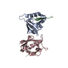
Mass: 22.990 Da / Num. of mol.: 1 / Source method: obtained synthetically / Formula: Na
#4: Water ChemComp-HOH / water


Mass: 18.015 Da / Num. of mol.: 132 / Source method: isolated from a natural source / Formula: H2O
Has ligand of interestN

-
Experimental details

-
Experiment

ExperimentMethod: X-RAY DIFFRACTION / Number of used crystals: 1

-
Sample preparation

CrystalDensity Matthews: 2.35 Å3/Da / Density % sol: 47.6 %
Crystal growTemperature: 277.15 K / Method: vapor diffusion, sitting drop / Details: 20% w/v PEG 3350, 0.2 M NaI at pH 7;

-
Data collection

DiffractionMean temperature: 100 K / Serial crystal experiment: N
Diffraction sourceSource: SYNCHROTRON / Site: SOLEIL / Beamline: PROXIMA 1 / Wavelength: 0.9786 Å
DetectorType: DECTRIS EIGER X 16M / Detector: PIXEL / Date: Apr 13, 2018
RadiationProtocol: SINGLE WAVELENGTH / Monochromatic (M) / Laue (L): M / Scattering type: x-ray
Radiation wavelengthWavelength: 0.9786 Å / Relative weight: 1
ReflectionResolution: 1.87→31.7 Å / Num. obs: 23304 / % possible obs: 99.17 % / Redundancy: 12.3 % / Biso Wilson estimate: 33.51 Å2 / CC1/2: 0.999 / Net I/σ(I): 23.94
Reflection shellResolution: 2.1→2.175 Å / Num. unique obs: 1630 / CC1/2: 0.973

-
Processing

Software
NameVersionClassification
PHENIX1.20.1_4487refinement
XDSdata reduction
Aimlessdata scaling
PHASERphasing
RefinementMethod to determine structure: MOLECULAR REPLACEMENT / Resolution: 1.87→31.7 Å / SU ML: 0.2741 / Cross valid method: FREE R-VALUE / σ(F): 1.33 / Phase error: 25.1486
Stereochemistry target values: GeoStd + Monomer Library + CDL v1.2
RfactorNum. reflection% reflection
Rfree0.2488 1164 5 %
Rwork0.1983 22130 -
obs0.2008 23294 98.73 %
Solvent computationShrinkage radii: 0.9 Å / VDW probe radii: 1.1 Å / Solvent model: FLAT BULK SOLVENT MODEL
Displacement parametersBiso mean: 45.16 Å2
Refinement stepCycle: LAST / Resolution: 1.87→31.7 Å
ProteinNucleic acidLigandSolventTotal
Num. atoms1624 0 1 132 1757
Refine LS restraints
Refine-IDTypeDev idealNumber
X-RAY DIFFRACTIONf_bond_d0.01211651
X-RAY DIFFRACTIONf_angle_d1.93342223
X-RAY DIFFRACTIONf_chiral_restr0.0818252
X-RAY DIFFRACTIONf_plane_restr0.0222297
X-RAY DIFFRACTIONf_dihedral_angle_d8.0565228
Refine LS restraints NCSType: Torsion NCS / Rms dev position: 1.03802942964 Å
LS refinement shell
Resolution (Å)Rfactor RfreeNum. reflection RfreeRfactor RworkNum. reflection RworkRefine-ID% reflection obs (%)
1.87-1.960.33841360.29542584X-RAY DIFFRACTION94.48
1.96-2.060.26541430.242715X-RAY DIFFRACTION99.69
2.06-2.190.31421440.23392752X-RAY DIFFRACTION99.97
2.19-2.360.25761450.21392751X-RAY DIFFRACTION99.86
2.36-2.60.24371460.21232770X-RAY DIFFRACTION99.73
2.6-2.970.31381470.21392791X-RAY DIFFRACTION99.59
2.97-3.740.23921480.19072820X-RAY DIFFRACTION98.97
3.74-31.70.21151550.17172947X-RAY DIFFRACTION97.64
Refinement TLS params.

Method: refined / Refine-ID: X-RAY DIFFRACTION

IDL112)L122)L132)L222)L232)L332)S11 (Å °)S12 (Å °)S13 (Å °)S21 (Å °)S22 (Å °)S23 (Å °)S31 (Å °)S32 (Å °)S33 (Å °)T112)T122)T132)T222)T232)T332)Origin x (Å)Origin y (Å)Origin z (Å)
12.920218871661.05381096757-1.37485463873.25679323282-0.5446005234984.856675644930.116466325978-0.2497385741070.06863106830190.124169433172-0.0356137472072-0.1762298811750.04013086860990.389970958655-0.1038476079640.1892573068660.0380950871769-0.005270646354040.286750942995-0.01752162400150.207729802707-3.19723199623-34.901960309619.6796961596
22.56841746596-3.17853314476-1.357120845164.091196072871.025442416663.36988388881-0.132781786779-1.397235218550.4946244855760.6576010239110.2975253254450.1698016305220.05193216312720.331277664546-0.09159773891740.369227778034-0.004875318264480.08594076070190.426097020563-0.07342243935440.313096341643-12.516551592-26.583970531131.0747834599
34.186846237640.470260576050.121534067873.0089145938-0.80405399093.275949387320.01002201020360.3020772167030.358379270668-0.1199101885730.09030122126620.0168917518566-0.168195483485-0.0735387280748-0.08465922650940.2313396940350.03693473708060.03222972462470.2427063189820.0657325389640.265301628636-0.755786015821-23.7424528572-3.73112156731
48.91699496071-1.07580879638-2.098318034147.351813535451.999325751191.912042412120.3407828117661.030785922460.323402505868-0.697935913821-0.147533980130.0687849861324-0.610305105397-0.669540060412-0.1045917998910.4962477557810.0879027626347-0.01684476972020.5340930422750.1592469473920.356017646779-6.05158382922-22.416790078-15.0513431482
Refinement TLS group

Refine-ID: X-RAY DIFFRACTION

IDRefine TLS-IDSelection detailsAuth asym-IDLabel asym-IDAuth seq-IDLabel seq-ID
11(chain 'A' and resid 5 through 97)AA5 - 971 - 93
22(chain 'B' and resid 1 through 11)BB1 - 111 - 11
33(chain 'C' and resid 6 through 97)CC6 - 971 - 92
44(chain 'D' and resid 6 through 11)DD6 - 111 - 6

+
About Yorodumi

-
News

-
Feb 9, 2022. New format data for meta-information of EMDB entries

New format data for meta-information of EMDB entries

  • Version 3 of the EMDB header file is now the official format.
  • The previous official version 1.9 will be removed from the archive.

Related info.:EMDB header

External links:wwPDB to switch to version 3 of the EMDB data model

-
Aug 12, 2020. Covid-19 info

Covid-19 info

URL: https://pdbjlvh1.pdbj.org/emnavi/covid19.php

New page: Covid-19 featured information page in EM Navigator.

Related info.:Covid-19 info / Mar 5, 2020. Novel coronavirus structure data

+
Mar 5, 2020. Novel coronavirus structure data

Novel coronavirus structure data

Related info.:Yorodumi Speices / Aug 12, 2020. Covid-19 info

External links:COVID-19 featured content - PDBj / Molecule of the Month (242):Coronavirus Proteases

+
Jan 31, 2019. EMDB accession codes are about to change! (news from PDBe EMDB page)

EMDB accession codes are about to change! (news from PDBe EMDB page)

  • The allocation of 4 digits for EMDB accession codes will soon come to an end. Whilst these codes will remain in use, new EMDB accession codes will include an additional digit and will expand incrementally as the available range of codes is exhausted. The current 4-digit format prefixed with “EMD-” (i.e. EMD-XXXX) will advance to a 5-digit format (i.e. EMD-XXXXX), and so on. It is currently estimated that the 4-digit codes will be depleted around Spring 2019, at which point the 5-digit format will come into force.
  • The EM Navigator/Yorodumi systems omit the EMD- prefix.

Related info.:Q: What is EMD? / ID/Accession-code notation in Yorodumi/EM Navigator

External links:EMDB Accession Codes are Changing Soon! / Contact to PDBj

+
Jul 12, 2017. Major update of PDB

Major update of PDB

  • wwPDB released updated PDB data conforming to the new PDBx/mmCIF dictionary.
  • This is a major update changing the version number from 4 to 5, and with Remediation, in which all the entries are updated.
  • In this update, many items about electron microscopy experimental information are reorganized (e.g. em_software).
  • Now, EM Navigator and Yorodumi are based on the updated data.

External links:wwPDB Remediation / Enriched Model Files Conforming to OneDep Data Standards Now Available in the PDB FTP Archive

-
Yorodumi

Thousand views of thousand structures

  • Yorodumi is a browser for structure data from EMDB, PDB, SASBDB, etc.
  • This page is also the successor to EM Navigator detail page, and also detail information page/front-end page for Omokage search.
  • The word "yorodu" (or yorozu) is an old Japanese word meaning "ten thousand". "mi" (miru) is to see.

Related info.:EMDB / PDB / SASBDB / Comparison of 3 databanks / Yorodumi Search / Aug 31, 2016. New EM Navigator & Yorodumi / Yorodumi Papers / Jmol/JSmol / Function and homology information / Changes in new EM Navigator and Yorodumi

Read more