 Movie
Movie Controller

[English] 日本語
 Yorodumi
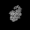
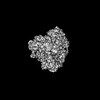
- PDB-8ki6: Structure of tomato spotted wilt virus L protein binding to Ribavirin -
+ Open data

- Basic information
| Entry | Database: PDB / ID: 8ki6 | |||||||||||||||||||||||||||||||||
|---|---|---|---|---|---|---|---|---|---|---|---|---|---|---|---|---|---|---|---|---|---|---|---|---|---|---|---|---|---|---|---|---|---|---|
| Title | Structure of tomato spotted wilt virus L protein binding to Ribavirin | |||||||||||||||||||||||||||||||||
 Components Components | RNA-directed RNA polymerase L | |||||||||||||||||||||||||||||||||
 Keywords Keywords | VIRAL PROTEIN / tomato spotted wilt virus / L protein | |||||||||||||||||||||||||||||||||
| Function / homology |  Function and homology information Function and homology informationRNA-directed RNA polymerase / viral RNA genome replication / RNA-directed RNA polymerase activity / DNA-templated transcription Similarity search - Function | |||||||||||||||||||||||||||||||||
| Biological species |  Orthotospovirus tomatomaculae Orthotospovirus tomatomaculae | |||||||||||||||||||||||||||||||||
| Method | ELECTRON MICROSCOPY / single particle reconstruction / cryo EM / Resolution: 3.4 Å | |||||||||||||||||||||||||||||||||
 Authors Authors | Cao, L. / Wang, X. | |||||||||||||||||||||||||||||||||
| Funding support | 1items
| |||||||||||||||||||||||||||||||||
 Citation Citation | Journal: Nat Plants / Year: 2025 Title: Structural basis for the activation of plant bunyavirus replication machinery and its dual-targeted inhibition by ribavirin. Authors: Jia Li / Lei Cao / Yaqian Zhao / Jinghan Shen / Lei Wang / Mingfeng Feng / Min Zhu / Yonghao Ye / Richard Kormelink / Xiaorong Tao / Xiangxi Wang /   Abstract: Despite the discovery of plant viruses as a new class of pathogens over a century ago, the structure of plant virus replication machinery and antiviral pesticide remains lacking. Here we report five ...Despite the discovery of plant viruses as a new class of pathogens over a century ago, the structure of plant virus replication machinery and antiviral pesticide remains lacking. Here we report five cryogenic electron microscopy structures of a ~330-kDa RNA-dependent RNA polymerase (RdRp) from a devastating plant bunyavirus, tomato spotted wilt orthotospovirus (TSWV), including the apo, viral-RNA-bound, base analogue ribavirin-bound and ribavirin-triphosphate-bound states. They reveal that a flexible loop of RdRp's motif F functions as 'sensor' to perceive viral RNA and further acts as an 'adaptor' to promote the formation of a complete catalytic centre. A ten-base RNA 'hook' structure is sufficient to trigger major conformational changes and activate RdRp. Chemical screening showed that ribavirin is effective against TSWV, and structural data revealed that ribavirin disrupts both hook-binding and catalytic core formation, locking polymerase in its inactive state. This work provides structural insights into the mechanisms of plant bunyavirus RdRp activation and its dual-targeted site inhibition, facilitating the development of pesticides against plant viruses. | |||||||||||||||||||||||||||||||||
| History |
|
- Structure visualization
| Structure viewer | Molecule: MolmilJmol/JSmol |
|---|
- Downloads & links
-Download
| PDBx/mmCIF format | 8ki6.cif.gz | 298.1 KB | Display | PDBx/mmCIF format |
|---|---|---|---|---|
| PDB format | pdb8ki6.ent.gz | 230.6 KB | Display | PDB format |
| PDBx/mmJSON format | 8ki6.json.gz | Tree view | PDBx/mmJSON format | |
| Others |  Other downloads Other downloads |
-Validation report
| Arichive directory | https://data.pdbj.org/pub/pdb/validation_reports/ki/8ki6ftp://data.pdbj.org/pub/pdb/validation_reports/ki/8ki6 | HTTPS FTP |
|---|
-Related structure data
| Related structure data | 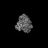 37252MC 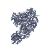 8ki7C 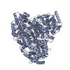 8ki8C  8ki9C 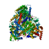 8kiaC 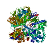 9j8vC M: map data used to model this data C: citing same article ( |
|---|---|
| Similar structure data |

-Links
 PDBj
PDBj- Assembly
| Deposited unit | 
|
|---|---|
| 1 |
|
-Components
| #1: Protein | Mass: 204262.906 Da / Num. of mol.: 1 Source method: isolated from a genetically manipulated source Source: (gene. exp.) Orthotospovirus tomatomaculaeProduction host: Spodoptera frugiperda multiple nucleopolyhedrovirusReferences: UniProt: A0A7G8JUQ9, RNA-directed RNA polymerase | ||||
|---|---|---|---|---|---|
| #2: Chemical | 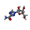  Mass: 244.205 Da / Num. of mol.: 2 / Source method: obtained synthetically / Formula: C8H12N4O5 / Feature type: SUBJECT OF INVESTIGATION Mass: 244.205 Da / Num. of mol.: 2 / Source method: obtained synthetically / Formula: C8H12N4O5 / Feature type: SUBJECT OF INVESTIGATIONHas ligand of interest | Y | Has protein modification | N | |
-Experimental details
-Experiment
| Experiment | Method: ELECTRON MICROSCOPY |
|---|---|
| EM experiment | Aggregation state: PARTICLE / 3D reconstruction method: single particle reconstruction |
- Sample preparation
| Component | Name: Structure of tomato spotted wilt virus L protein binding to Ribavirin Type: COMPLEX / Entity ID: #1 / Source: RECOMBINANT |
|---|---|
| Source (natural) | Organism: Orthotospovirus tomatomaculae |
| Source (recombinant) | Organism:   Spodoptera frugiperda (fall armyworm) Spodoptera frugiperda (fall armyworm) |
| Buffer solution | pH: 7.5 |
| Specimen | Embedding applied: NO / Shadowing applied: NO / Staining applied: NO / Vitrification applied: YES |
| Vitrification | Cryogen name: ETHANE |
- Electron microscopy imaging
| Microscopy | Model: FEI TITAN |
|---|---|
| Electron gun | Electron source:  FIELD EMISSION GUN / Accelerating voltage: 300 kV / Illumination mode: FLOOD BEAM FIELD EMISSION GUN / Accelerating voltage: 300 kV / Illumination mode: FLOOD BEAM |
| Electron lens | Mode: BRIGHT FIELD / Nominal defocus max: 1800 nm / Nominal defocus min: 1200 nm |
| Image recording | Electron dose: 60 e/Å2 / Film or detector model: GATAN K2 QUANTUM (4k x 4k) |
- Processing
| EM software | Name: PHENIX / Category: model refinement | ||||||||||||||||||||||||
|---|---|---|---|---|---|---|---|---|---|---|---|---|---|---|---|---|---|---|---|---|---|---|---|---|---|
| CTF correction | Type: PHASE FLIPPING AND AMPLITUDE CORRECTION | ||||||||||||||||||||||||
| 3D reconstruction | Resolution: 3.4 Å / Resolution method: FSC 0.143 CUT-OFF / Num. of particles: 348921 / Symmetry type: POINT | ||||||||||||||||||||||||
| Refine LS restraints |
|
