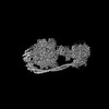
 Movie
Movie Controller

[English] 日本語
 Yorodumi
- PDB-8khf: Structure of the human ATP synthase bound to bedaquiline (membran... -
+ Open data

- Basic information
| Entry | Database: PDB / ID: 8khf | ||||||||||||
|---|---|---|---|---|---|---|---|---|---|---|---|---|---|
| Title | Structure of the human ATP synthase bound to bedaquiline (membrane domain) | ||||||||||||
 Components Components |
| ||||||||||||
 Keywords Keywords | MEMBRANE PROTEIN / ATP synthase / Human / cryo-EM | ||||||||||||
| Function / homology |  Function and homology information Function and homology informationFormation of ATP by chemiosmotic coupling / Cristae formation / ATP biosynthetic process / mitochondrial proton-transporting ATP synthase complex assembly / Mitochondrial protein import / Mitochondrial translation termination / oxidative phosphorylation / proton channel activity / response to copper ion / proton transmembrane transporter activity ...Formation of ATP by chemiosmotic coupling / Cristae formation / ATP biosynthetic process / mitochondrial proton-transporting ATP synthase complex assembly / Mitochondrial protein import / Mitochondrial translation termination / oxidative phosphorylation / proton channel activity / response to copper ion / proton transmembrane transporter activity / proton motive force-driven ATP synthesis / proton-transporting two-sector ATPase complex, proton-transporting domain / proton motive force-driven mitochondrial ATP synthesis / response to hyperoxia / proton-transporting ATP synthase complex / proton-transporting ATP synthase activity, rotational mechanism / proton transmembrane transport / substantia nigra development / Mitochondrial protein degradation / aerobic respiration / mitochondrial inner membrane / mitochondrial matrix / hydrolase activity / lipid binding / protein-containing complex binding / structural molecule activity / mitochondrion / RNA binding / membrane / nucleus Similarity search - Function | ||||||||||||
| Biological species |  Homo sapiens (human) Homo sapiens (human) | ||||||||||||
| Method | ELECTRON MICROSCOPY / single particle reconstruction / cryo EM / Resolution: 3.13 Å | ||||||||||||
 Authors Authors | Lai, Y. / Zhang, Y. | ||||||||||||
| Funding support |  China, 3items China, 3items
| ||||||||||||
 Citation Citation | Journal: Nature / Year: 2024 Title: Inhibition of M. tuberculosis and human ATP synthase by BDQ and TBAJ-587. Authors: Yuying Zhang / Yuezheng Lai / Shan Zhou / Ting Ran / Yue Zhang / Ziqing Zhao / Ziyan Feng / Long Yu / Jinxu Xu / Kun Shi / Jianyun Wang / Yu Pang / Liang Li / Hongming Chen / Luke W Guddat / ...Authors: Yuying Zhang / Yuezheng Lai / Shan Zhou / Ting Ran / Yue Zhang / Ziqing Zhao / Ziyan Feng / Long Yu / Jinxu Xu / Kun Shi / Jianyun Wang / Yu Pang / Liang Li / Hongming Chen / Luke W Guddat / Yan Gao / Fengjiang Liu / Zihe Rao / Hongri Gong /  Abstract: Bedaquiline (BDQ), a first-in-class diarylquinoline anti-tuberculosis drug, and its analogue, TBAJ-587, prevent the growth and proliferation of Mycobacterium tuberculosis by inhibiting ATP synthase. ...Bedaquiline (BDQ), a first-in-class diarylquinoline anti-tuberculosis drug, and its analogue, TBAJ-587, prevent the growth and proliferation of Mycobacterium tuberculosis by inhibiting ATP synthase. However, BDQ also inhibits human ATP synthase. At present, how these compounds interact with either M. tuberculosis ATP synthase or human ATP synthase is unclear. Here we present cryogenic electron microscopy structures of M. tuberculosis ATP synthase with and without BDQ and TBAJ-587 bound, and human ATP synthase bound to BDQ. The two inhibitors interact with subunit a and the c-ring at the leading site, c-only sites and lagging site in M. tuberculosis ATP synthase, showing that BDQ and TBAJ-587 have similar modes of action. The quinolinyl and dimethylamino units of the compounds make extensive contacts with the protein. The structure of human ATP synthase in complex with BDQ reveals that the BDQ-binding site is similar to that observed for the leading site in M. tuberculosis ATP synthase, and that the quinolinyl unit also interacts extensively with the human enzyme. This study will improve researchers' understanding of the similarities and differences between human ATP synthase and M. tuberculosis ATP synthase in terms of the mode of BDQ binding, and will allow the rational design of novel diarylquinolines as anti-tuberculosis drugs. | ||||||||||||
| History |
|
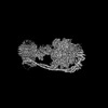
- Structure visualization
| Structure viewer | Molecule: MolmilJmol/JSmol |
|---|
- Downloads & links
-Download
| PDBx/mmCIF format | 8khf.cif.gz | 283.3 KB | Display | PDBx/mmCIF format |
|---|---|---|---|---|
| PDB format | pdb8khf.ent.gz | 224.6 KB | Display | PDB format |
| PDBx/mmJSON format | 8khf.json.gz | Tree view | PDBx/mmJSON format | |
| Others |  Other downloads Other downloads |
-Validation report
| Arichive directory | https://data.pdbj.org/pub/pdb/validation_reports/kh/8khfftp://data.pdbj.org/pub/pdb/validation_reports/kh/8khf | HTTPS FTP |
|---|
-Related structure data
| Related structure data |  37243MC 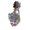 8j0sC 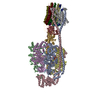 8j0tC 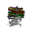 8j57C  8j58C 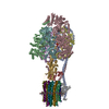 8jr0C  8jr1C  8ki3C M: map data used to model this data C: citing same article ( |
|---|---|
| Similar structure data |


-Links
 PDBj
PDBj

- Assembly
| Deposited unit | 
|
|---|---|
| 1 |
|
-Components
-ATP synthase F(0) complex subunit ... , 2 types, 9 molecules 12345678K
| #1: Protein | Mass: 7610.954 Da / Num. of mol.: 8 / Source method: isolated from a natural source / Source: (natural) Homo sapiens (human) / References: UniProt: P05496#5: Protein | | Mass: 24658.586 Da / Num. of mol.: 1 / Source method: isolated from a natural source / Source: (natural) Homo sapiens (human) / References: UniProt: P24539 |
|---|
-ATP synthase subunit ... , 9 types, 9 molecules GHIMNPRST
| #2: Protein | Mass: 30207.752 Da / Num. of mol.: 1 / Source method: isolated from a natural source / Source: (natural) Homo sapiens (human) / References: UniProt: P36542 |
|---|---|
| #3: Protein | Mass: 15029.817 Da / Num. of mol.: 1 / Source method: isolated from a natural source / Source: (natural) Homo sapiens (human) / References: UniProt: P30049 |
| #4: Protein | Mass: 5790.779 Da / Num. of mol.: 1 / Source method: isolated from a natural source / Source: (natural) Homo sapiens (human) / References: UniProt: P56381 |
| #6: Protein | Mass: 18383.982 Da / Num. of mol.: 1 / Source method: isolated from a natural source / Source: (natural) Homo sapiens (human) / References: UniProt: O75947 |
| #7: Protein | Mass: 24833.102 Da / Num. of mol.: 1 / Source method: isolated from a natural source / Source: (natural) Homo sapiens (human) / References: UniProt: P00846 |
| #8: Protein | Mass: 6673.053 Da / Num. of mol.: 1 / Source method: isolated from a natural source / Source: (natural) Homo sapiens (human) / References: UniProt: P56378 |
| #10: Protein | Mass: 10804.686 Da / Num. of mol.: 1 / Source method: isolated from a natural source / Source: (natural) Homo sapiens (human) / References: UniProt: P56134 |
| #11: Protein | Mass: 11309.226 Da / Num. of mol.: 1 / Source method: isolated from a natural source / Source: (natural) Homo sapiens (human) / References: UniProt: O75964 |
| #12: Protein | Mass: 7947.215 Da / Num. of mol.: 1 / Source method: isolated from a natural source / Source: (natural) Homo sapiens (human) / References: UniProt: P56385 |
-Protein / Non-polymers , 2 types, 2 molecules Q
| #13: Chemical | ChemComp-BQ1 /  Mass: 555.505 Da / Num. of mol.: 1 / Source method: obtained synthetically / Formula: C32H31BrN2O2 / Feature type: SUBJECT OF INVESTIGATION / Comment: antibiotic*YM Mass: 555.505 Da / Num. of mol.: 1 / Source method: obtained synthetically / Formula: C32H31BrN2O2 / Feature type: SUBJECT OF INVESTIGATION / Comment: antibiotic*YM |
|---|---|
| #9: Protein | Mass: 8000.634 Da / Num. of mol.: 1 / Source method: isolated from a natural source / Source: (natural) Homo sapiens (human) / References: UniProt: P03928 |
-Details
| Has ligand of interest | Y |
|---|
-Experimental details
-Experiment
| Experiment | Method: ELECTRON MICROSCOPY |
|---|---|
| EM experiment | Aggregation state: PARTICLE / 3D reconstruction method: single particle reconstruction |
- Sample preparation
| Component | Name: human ATP synthase / Type: COMPLEX / Entity ID: #1-#12 / Source: NATURAL |
|---|---|
| Source (natural) | Organism: Homo sapiens (human) |
| Source (recombinant) | Organism: Homo sapiens (human) |
| Buffer solution | pH: 7.4 |
| Specimen | Embedding applied: NO / Shadowing applied: NO / Staining applied: NO / Vitrification applied: YES |
| Vitrification | Cryogen name: ETHANE |
- Electron microscopy imaging
| Experimental equipment |  Model: Titan Krios / Image courtesy: FEI Company |
|---|---|
| Microscopy | Model: FEI TITAN KRIOS |
| Electron gun | Electron source:  FIELD EMISSION GUN / Accelerating voltage: 300 kV / Illumination mode: FLOOD BEAM FIELD EMISSION GUN / Accelerating voltage: 300 kV / Illumination mode: FLOOD BEAM |
| Electron lens | Mode: BRIGHT FIELD / Nominal defocus max: 2400 nm / Nominal defocus min: 1200 nm |
| Image recording | Electron dose: 50 e/Å2 / Film or detector model: FEI FALCON IV (4k x 4k) |
- Processing
| CTF correction | Type: PHASE FLIPPING AND AMPLITUDE CORRECTION |
|---|---|
| 3D reconstruction | Resolution: 3.13 Å / Resolution method: FSC 0.143 CUT-OFF / Num. of particles: 84037 / Symmetry type: POINT |
