| 登録構造単位 | C: antibody heavy chain
D: antibody light chain
J: T-cell immunoreceptor with Ig and ITIM domains
A: antibody heavy chain
B: antibody light chain
E: T-cell immunoreceptor with Ig and ITIM domains
F: antibody heavy chain
G: antibody light chain
H: T-cell immunoreceptor with Ig and ITIM domains
I: antibody heavy chain
K: antibody light chain
L: T-cell immunoreceptor with Ig and ITIM domains
| 分子量 (理論値) | 分子数 |
|---|
| 合計 (水以外) | 239,387 | 12 |
|---|
| ポリマ- | 239,387 | 12 |
|---|
| 非ポリマー | 0 | 0 |
|---|
| 水 | 14,736 | 818 |
|---|
|
|---|
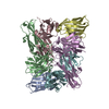
| 1 | C: antibody heavy chain
D: antibody light chain
J: T-cell immunoreceptor with Ig and ITIM domains
 - 登録者が定義した集合体
- 根拠:
 gel filtration, chain A, B, E chain I, K, L chain C, D, J chain F, G, H gel filtration, chain A, B, E chain I, K, L chain C, D, J chain F, G, H - 59.8 kDa, 3 ポリマー
 Omokage検索でこの集合体の類似形状データを探す (詳細) Omokage検索でこの集合体の類似形状データを探す (詳細)
| 分子量 (理論値) | 分子数 |
|---|
| 合計 (水以外) | 59,847 | 3 |
|---|
| ポリマ- | 59,847 | 3 |
|---|
| 非ポリマー | 0 | 0 |
|---|
| 水 | 54 | 3 |
|---|
| タイプ | 名称 | 対称操作 | 数 |
|---|
| identity operation | 1_555 | x,y,z | 1 |
|
|---|
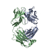
| 2 | A: antibody heavy chain
B: antibody light chain
E: T-cell immunoreceptor with Ig and ITIM domains
| 分子量 (理論値) | 分子数 |
|---|
| 合計 (水以外) | 59,847 | 3 |
|---|
| ポリマ- | 59,847 | 3 |
|---|
| 非ポリマー | 0 | 0 |
|---|
| 水 | 54 | 3 |
|---|
| タイプ | 名称 | 対称操作 | 数 |
|---|
| identity operation | 1_555 | x,y,z | 1 |
|
|---|
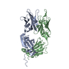
| 3 | F: antibody heavy chain
G: antibody light chain
H: T-cell immunoreceptor with Ig and ITIM domains
| 分子量 (理論値) | 分子数 |
|---|
| 合計 (水以外) | 59,847 | 3 |
|---|
| ポリマ- | 59,847 | 3 |
|---|
| 非ポリマー | 0 | 0 |
|---|
| 水 | 54 | 3 |
|---|
| タイプ | 名称 | 対称操作 | 数 |
|---|
| identity operation | 1_555 | x,y,z | 1 |
|
|---|
| 4 | I: antibody heavy chain
K: antibody light chain
L: T-cell immunoreceptor with Ig and ITIM domains
| 分子量 (理論値) | 分子数 |
|---|
| 合計 (水以外) | 59,847 | 3 |
|---|
| ポリマ- | 59,847 | 3 |
|---|
| 非ポリマー | 0 | 0 |
|---|
| 水 | 54 | 3 |
|---|
| タイプ | 名称 | 対称操作 | 数 |
|---|
| identity operation | 1_555 | x,y,z | 1 |
|
|---|
| 単位格子 | | Length a, b, c (Å) | 135.530, 138.310, 196.800 |
|---|
| Angle α, β, γ (deg.) | 90.00, 90.00, 90.00 |
|---|
| Int Tables number | 18 |
|---|
| Space group name H-M | P22121 |
|---|
|
|---|
| Components on special symmetry positions | | ID | モデル | 要素 |
|---|
| 1 | 1 | A-378- HOH |
|
|---|
 ムービー
ムービー コントローラー

 データを開く
 基本情報
基本情報 要素
要素 キーワード
キーワード 機能・相同性情報
 Homo sapiens (ヒト)
 データ登録者
 引用
引用 構造の表示
構造の表示 ダウンロードとリンク
 その他のダウンロード
 PDBj
PDBj

 集合体
集合体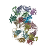


 試料調製
試料調製 / ビームライン: BL45XU / 波長: 1 Å
 解析
解析