National Natural Science Foundation of China (NSFC)
中国
引用
ジャーナル: Nat Commun / 年: 2024 タイトル: Self-assembled superstructure alleviates air-water interface effect in cryo-EM. 著者: Liming Zheng / Jie Xu / Weihua Wang / Xiaoyin Gao / Chao Zhao / Weijun Guo / Luzhao Sun / Hang Cheng / Fanhao Meng / Buhang Chen / Weiyu Sun / Xia Jia / Xiong Zhou / Kai Wu / Zhongfan Liu / ...著者: Liming Zheng / Jie Xu / Weihua Wang / Xiaoyin Gao / Chao Zhao / Weijun Guo / Luzhao Sun / Hang Cheng / Fanhao Meng / Buhang Chen / Weiyu Sun / Xia Jia / Xiong Zhou / Kai Wu / Zhongfan Liu / Feng Ding / Nan Liu / Hong-Wei Wang / Hailin Peng / 要旨: Cryo-electron microscopy (cryo-EM) has been widely used to reveal the structures of proteins at atomic resolution. One key challenge is that almost all proteins are predominantly adsorbed to the air- ...Cryo-electron microscopy (cryo-EM) has been widely used to reveal the structures of proteins at atomic resolution. One key challenge is that almost all proteins are predominantly adsorbed to the air-water interface during standard cryo-EM specimen preparation. The interaction of proteins with air-water interface will significantly impede the success of reconstruction and achievable resolution. Here, we highlight the critical role of impenetrable surfactant monolayers in passivating the air-water interface problems, and develop a robust effective method for high-resolution cryo-EM analysis, by using the superstructure GSAMs which comprises surfactant self-assembled monolayers (SAMs) and graphene membrane. The GSAMs works well in enriching the orientations and improving particle utilization ratio of multiple proteins, facilitating the 3.3-Å resolution reconstruction of a 100-kDa protein complex (ACE2-RBD), which shows strong preferential orientation using traditional specimen preparation protocol. Additionally, we demonstrate that GSAMs enables the successful determinations of small proteins (<100 kDa) at near-atomic resolution. This study expands the understanding of SAMs and provides a key to better control the interaction of protein with air-water interface.
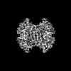
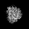
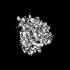
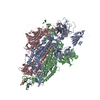
Spikeglycoprotein / S glycoprotein / E2 / Peplomer protein
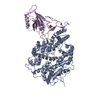
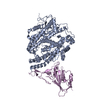
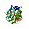
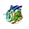
分子量: 138842.375 Da / 分子数: 3 / 由来タイプ: 組換発現 詳細: For issue of poor atom inclusion, the depositor stated 'we have tried our best to fit the coordinates in the density map of spike via rigid-body dock. However, the flexibility of RBD makes it ...詳細: For issue of poor atom inclusion, the depositor stated 'we have tried our best to fit the coordinates in the density map of spike via rigid-body dock. However, the flexibility of RBD makes it hard to fit, which seems to be general for spike proteins.' 由来: (組換発現) Severe acute respiratory syndrome coronavirus 2 (ウイルス) 遺伝子: S, 2 / 発現宿主: Homo sapiens (ヒト) / 参照: UniProt: P0DTC2
Has protein modification
Y
-
実験情報
-
実験
実験
手法: 電子顕微鏡法
EM実験
試料の集合状態: PARTICLE / 3次元再構成法: 単粒子再構成法
-
試料調製
構成要素
名称: SARS-CoV-2 Delta variant spike protein / タイプ: COMPLEX / 詳細: on graphene membranes / Entity ID: all / 由来: RECOMBINANT
由来(天然)
生物種: Severe acute respiratory syndrome coronavirus 2 (ウイルス)
由来(組換発現)
生物種: Homo sapiens (ヒト)
緩衝液
pH: 7.8
試料
濃度: 0.25 mg/ml / 包埋: NO / シャドウイング: NO / 染色: NO / 凍結: YES
 ムービー
ムービー コントローラー


 データを開く
 基本情報
基本情報 要素
要素 キーワード
キーワード 機能・相同性情報
 Severe acute respiratory syndrome coronavirus 2 (ウイルス)
 データ登録者
 中国, 1件
  引用
引用 構造の表示
構造の表示 ダウンロードとリンク
 その他のダウンロード


 PDBj
PDBj


 集合体
集合体
 Homo sapiens (ヒト) / 参照: UniProt: P0DTC2
 試料調製
試料調製 電子顕微鏡撮影
 FIELD EMISSION GUN / 加速電圧: 300 kV / 照射モード: FLOOD BEAM
 解析
解析