 Movie
Movie Controller

[English] 日本語
 Yorodumi
- PDB-8hjp: Crystal structure of glycosyltransferase SgUGT94-289-3 in complex... -
+ Open data

- Basic information
| Entry | Database: PDB / ID: 8hjp | |||||||||||||||
|---|---|---|---|---|---|---|---|---|---|---|---|---|---|---|---|---|
| Title | Crystal structure of glycosyltransferase SgUGT94-289-3 in complex with UDP state 1 | |||||||||||||||
 Components Components | glycosyltranseferease | |||||||||||||||
 Keywords Keywords | TRANSFERASE / mogroside / UGT / Siraitia grosvenorii | |||||||||||||||
| Function / homology | Chem-POG / URIDINE-5'-DIPHOSPHATE Function and homology information Function and homology information | |||||||||||||||
| Biological species |  Siraitia grosvenorii (monk fruit) Siraitia grosvenorii (monk fruit) | |||||||||||||||
| Method |  X-RAY DIFFRACTION / SYNCHROTRON / MOLECULAR REPLACEMENT / Resolution: 2.2 Å X-RAY DIFFRACTION / SYNCHROTRON / MOLECULAR REPLACEMENT / Resolution: 2.2 Å | |||||||||||||||
 Authors Authors | Li, M. / Zhang, S. / Cui, S. | |||||||||||||||
| Funding support |  China, China,  United Kingdom, 4items United Kingdom, 4items
| |||||||||||||||
 Citation Citation | Journal: Nat Commun / Year: 2024 Title: Structural insights into the catalytic selectivity of glycosyltransferase SgUGT94-289-3 towards mogrosides. Authors: Cui, S. / Zhang, S. / Wang, N. / Su, X. / Luo, Z. / Ma, X. / Li, M. | |||||||||||||||
| History |
|
- Structure visualization
| Structure viewer | Molecule: MolmilJmol/JSmol |
|---|
- Downloads & links
-Download
| PDBx/mmCIF format | 8hjp.cif.gz | 193.3 KB | Display | PDBx/mmCIF format |
|---|---|---|---|---|
| PDB format | pdb8hjp.ent.gz | 148.6 KB | Display | PDB format |
| PDBx/mmJSON format | 8hjp.json.gz | Tree view | PDBx/mmJSON format | |
| Others |  Other downloads Other downloads |
-Validation report
| Arichive directory | https://data.pdbj.org/pub/pdb/validation_reports/hj/8hjpftp://data.pdbj.org/pub/pdb/validation_reports/hj/8hjp | HTTPS FTP |
|---|
-Related structure data
| Related structure data | 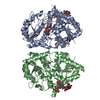 8hjfC 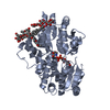 8hjgC 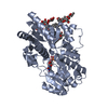 8hjhC 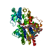 8hjkC 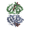 8hjlC 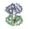 8hjnC  8hjoC 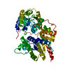 8hjqC 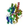 8j66C  6jtdS S: Starting model for refinement C: citing same article ( |
|---|---|
| Similar structure data |
-Links
 PDBj
PDBj


- Assembly
| Deposited unit | 
| ||||||||||||
|---|---|---|---|---|---|---|---|---|---|---|---|---|---|
| 1 |
| ||||||||||||
| 2 | 
| ||||||||||||
| Unit cell |
|
-Components
| #1: Protein | Mass: 51608.793 Da / Num. of mol.: 2 Source method: isolated from a genetically manipulated source Source: (gene. exp.) Siraitia grosvenorii (monk fruit) / Production host:  #2: Chemical | ChemComp-POG / ( |   Mass: 424.569 Da / Num. of mol.: 1 / Source method: obtained synthetically / Formula: C21H44O8 Mass: 424.569 Da / Num. of mol.: 1 / Source method: obtained synthetically / Formula: C21H44O8#3: Chemical |   Type: RNA linking / Mass: 404.161 Da / Num. of mol.: 2 / Source method: obtained synthetically / Formula: C9H14N2O12P2 / Feature type: SUBJECT OF INVESTIGATION / Comment: UDP*YM Type: RNA linking / Mass: 404.161 Da / Num. of mol.: 2 / Source method: obtained synthetically / Formula: C9H14N2O12P2 / Feature type: SUBJECT OF INVESTIGATION / Comment: UDP*YM#4: Water | ChemComp-HOH / |  Mass: 18.015 Da / Num. of mol.: 322 / Source method: isolated from a natural source / Formula: H2O Mass: 18.015 Da / Num. of mol.: 322 / Source method: isolated from a natural source / Formula: H2OHas ligand of interest | Y | Has protein modification | Y | |
|---|
-Experimental details
-Experiment
| Experiment | Method: X-RAY DIFFRACTION / Number of used crystals: 1 |
|---|
- Sample preparation
| Crystal | Density Matthews: 2.18 Å3/Da / Density % sol: 43.56 % |
|---|---|
| Crystal grow | Temperature: 289.15 K / Method: vapor diffusion, hanging drop Details: Tris-HCl, polyethylene glycol 3350, NaCl, Polypropylene glycol P400. |
-Data collection
| Diffraction | Mean temperature: 100 K / Serial crystal experiment: N |
|---|---|
| Diffraction source | Source: SYNCHROTRON / Site: SSRF / Beamline: BL17U1 / Wavelength: 0.97918 Å |
| Detector | Type: DECTRIS EIGER X 16M / Detector: PIXEL / Date: Apr 16, 2020 |
| Radiation | Protocol: SINGLE WAVELENGTH / Monochromatic (M) / Laue (L): M / Scattering type: x-ray |
| Radiation wavelength | Wavelength: 0.97918 Å / Relative weight: 1 |
| Reflection | Resolution: 2.2→35.84 Å / Num. obs: 46304 / % possible obs: 99.65 % / Redundancy: 12.8 % / Biso Wilson estimate: 30.48 Å2 / CC1/2: 0.999 / Net I/σ(I): 15.91 |
| Reflection shell | Resolution: 2.2→2.279 Å / Num. unique obs: 4553 / CC1/2: 0.964 |
- Processing
| Software |
| ||||||||||||||||||||||||||||||||||||||||||||||||||||||||||||||||||||||||||||||||||||||||||||||||||||||||||||||||||||||||||||||
|---|---|---|---|---|---|---|---|---|---|---|---|---|---|---|---|---|---|---|---|---|---|---|---|---|---|---|---|---|---|---|---|---|---|---|---|---|---|---|---|---|---|---|---|---|---|---|---|---|---|---|---|---|---|---|---|---|---|---|---|---|---|---|---|---|---|---|---|---|---|---|---|---|---|---|---|---|---|---|---|---|---|---|---|---|---|---|---|---|---|---|---|---|---|---|---|---|---|---|---|---|---|---|---|---|---|---|---|---|---|---|---|---|---|---|---|---|---|---|---|---|---|---|---|---|---|---|---|
| Refinement | Method to determine structure: MOLECULAR REPLACEMENT Starting model: 6JTD Resolution: 2.2→35.84 Å / SU ML: 0.2836 / Cross valid method: FREE R-VALUE / σ(F): 1.34 / Phase error: 35.2437 Stereochemistry target values: GeoStd + Monomer Library + CDL v1.2
| ||||||||||||||||||||||||||||||||||||||||||||||||||||||||||||||||||||||||||||||||||||||||||||||||||||||||||||||||||||||||||||||
| Solvent computation | Shrinkage radii: 0.9 Å / VDW probe radii: 1.11 Å / Solvent model: FLAT BULK SOLVENT MODEL | ||||||||||||||||||||||||||||||||||||||||||||||||||||||||||||||||||||||||||||||||||||||||||||||||||||||||||||||||||||||||||||||
| Displacement parameters | Biso mean: 38.48 Å2 | ||||||||||||||||||||||||||||||||||||||||||||||||||||||||||||||||||||||||||||||||||||||||||||||||||||||||||||||||||||||||||||||
| Refinement step | Cycle: LAST / Resolution: 2.2→35.84 Å
| ||||||||||||||||||||||||||||||||||||||||||||||||||||||||||||||||||||||||||||||||||||||||||||||||||||||||||||||||||||||||||||||
| Refine LS restraints |
| ||||||||||||||||||||||||||||||||||||||||||||||||||||||||||||||||||||||||||||||||||||||||||||||||||||||||||||||||||||||||||||||
| LS refinement shell |
|
