 Movie
Movie Controller

[English] 日本語
 Yorodumi
- PDB-8hdn: Brucella melitensis 7-alpha-Hydroxysteroid Dehydrogenase mutant: ... -
+ Open data

- Basic information
| Entry | Database: PDB / ID: 8hdn | |||||||||
|---|---|---|---|---|---|---|---|---|---|---|
| Title | Brucella melitensis 7-alpha-Hydroxysteroid Dehydrogenase mutant: 1-53 truncation-I258M/K262T | |||||||||
 Components Components | 7-alpha-hydroxysteroid dehydrogenase | |||||||||
 Keywords Keywords | OXIDOREDUCTASE / SDR family / hydroxysteroid dehydrogenase | |||||||||
| Function / homology |  Function and homology information Function and homology information7alpha-hydroxysteroid dehydrogenase / cholate 7-alpha-dehydrogenase (NAD+) activity / monocarboxylic acid metabolic process / lipid metabolic process / nucleotide binding Similarity search - Function | |||||||||
| Biological species |  Brucella melitensis biotype 1 (bacteria) Brucella melitensis biotype 1 (bacteria) | |||||||||
| Method |  X-RAY DIFFRACTION / SYNCHROTRON / MOLECULAR REPLACEMENT / Resolution: 1.7 Å X-RAY DIFFRACTION / SYNCHROTRON / MOLECULAR REPLACEMENT / Resolution: 1.7 Å | |||||||||
 Authors Authors | Liu, Z.Y. / Zhang, R.Z. | |||||||||
| Funding support |  China, 2items China, 2items
| |||||||||
 Citation Citation | Journal: To Be Published Title: Structure of 7-alpha-hydroxysteroid dehydrogenase Authors: Liu, Z.Y. / Zhang, R.Z. | |||||||||
| History |
|
- Structure visualization
| Structure viewer | Molecule: MolmilJmol/JSmol |
|---|
- Downloads & links
-Download
| PDBx/mmCIF format | 8hdn.cif.gz | 199.3 KB | Display | PDBx/mmCIF format |
|---|---|---|---|---|
| PDB format | pdb8hdn.ent.gz | 156.6 KB | Display | PDB format |
| PDBx/mmJSON format | 8hdn.json.gz | Tree view | PDBx/mmJSON format | |
| Others |  Other downloads Other downloads |
-Validation report
| Arichive directory | https://data.pdbj.org/pub/pdb/validation_reports/hd/8hdnftp://data.pdbj.org/pub/pdb/validation_reports/hd/8hdn | HTTPS FTP |
|---|
-Related structure data
| Related structure data |  8hdeC 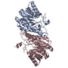 8hdiC 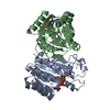 8hs4C 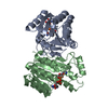 8hs5C 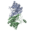 8hs6C 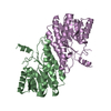 8hs9C 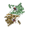 8hsaC 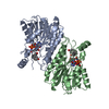 1fmcS 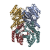 3gafS S: Starting model for refinement C: citing same article ( |
|---|---|
| Similar structure data |
-Links
 PDBj
PDBj


- Assembly
| Deposited unit | 
| ||||||||
|---|---|---|---|---|---|---|---|---|---|
| 1 | 
| ||||||||
| 2 | 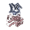
| ||||||||
| 3 | 
| ||||||||
| 4 | 
| ||||||||
| Unit cell |
|
-Components
| #1: Protein | Mass: 25872.635 Da / Num. of mol.: 4 / Fragment: 1-53 truncation Source method: isolated from a genetically manipulated source Source: (gene. exp.) Brucella melitensis biotype 1 (strain 16M / ATCC 23456 / NCTC 10094) (bacteria)Gene: BMEI0406 / Production host: References: UniProt: Q8YIN7, 7alpha-hydroxysteroid dehydrogenase #2: Water | ChemComp-HOH / |  Mass: 18.015 Da / Num. of mol.: 813 / Source method: isolated from a natural source / Formula: H2O Mass: 18.015 Da / Num. of mol.: 813 / Source method: isolated from a natural source / Formula: H2O |
|---|
-Experimental details
-Experiment
| Experiment | Method: X-RAY DIFFRACTION / Number of used crystals: 1 |
|---|
- Sample preparation
| Crystal | Density Matthews: 2.61 Å3/Da / Density % sol: 52.81 % |
|---|---|
| Crystal grow | Temperature: 289 K / Method: vapor diffusion, sitting drop / Details: 0.1M MIB, 10% PEG3350 |
-Data collection
| Diffraction | Mean temperature: 100 K / Serial crystal experiment: N |
|---|---|
| Diffraction source | Source: SYNCHROTRON / Site: SSRF / Beamline: BL17U1 / Wavelength: 0.987 Å |
| Detector | Type: DECTRIS EIGER X 16M / Detector: PIXEL / Date: Jul 12, 2021 |
| Radiation | Protocol: SINGLE WAVELENGTH / Monochromatic (M) / Laue (L): M / Scattering type: x-ray |
| Radiation wavelength | Wavelength: 0.987 Å / Relative weight: 1 |
| Reflection | Resolution: 1.7→44.13 Å / Num. obs: 113642 / % possible obs: 99.9 % / Redundancy: 9.9 % / Biso Wilson estimate: 12.68 Å2 / CC1/2: 0.996 / Rmerge(I) obs: 0.132 / Rpim(I) all: 0.044 / Rrim(I) all: 0.139 / Net I/σ(I): 10.9 |
| Reflection shell | Resolution: 1.7→1.763 Å / Rmerge(I) obs: 0.743 / Mean I/σ(I) obs: 10.9 / Num. unique obs: 11259 / CC1/2: 0.865 / Rpim(I) all: 0.334 / % possible all: 100 |
- Processing
| Software |
| ||||||||||||||||||||
|---|---|---|---|---|---|---|---|---|---|---|---|---|---|---|---|---|---|---|---|---|---|
| Refinement | Method to determine structure: MOLECULAR REPLACEMENT Starting model: 3GAF, 1FMC Resolution: 1.7→33.96 Å / SU ML: 0.15 / Cross valid method: FREE R-VALUE / σ(F): 1.97 / Phase error: 20.44 / Stereochemistry target values: ML
| ||||||||||||||||||||
| Solvent computation | Shrinkage radii: 0.9 Å / VDW probe radii: 1.1 Å / Solvent model: FLAT BULK SOLVENT MODEL | ||||||||||||||||||||
| Refinement step | Cycle: LAST / Resolution: 1.7→33.96 Å
| ||||||||||||||||||||
| Refine LS restraints | Type: f_bond_d / Dev ideal: 0.006 / Number: 7141 | ||||||||||||||||||||
| LS refinement shell | Resolution: 1.7→1.79 Å
|
