 Movie
Movie Controller

[English] 日本語
 Yorodumi
- PDB-8hdi: Brucella melitensis 7-alpha-Hydroxysteroid Dehydrogenase mutant: ... -
+ Open data

- Basic information
| Entry | Database: PDB / ID: 8hdi | |||||||||
|---|---|---|---|---|---|---|---|---|---|---|
| Title | Brucella melitensis 7-alpha-Hydroxysteroid Dehydrogenase mutant: 1-53 truncation/K262T | |||||||||
 Components Components | 7-alpha-hydroxysteroid dehydrogenase | |||||||||
 Keywords Keywords | OXIDOREDUCTASE / SDR family / hydroxysteroid dehydrogenase | |||||||||
| Function / homology |  Function and homology information Function and homology information7alpha-hydroxysteroid dehydrogenase / cholate 7-alpha-dehydrogenase (NAD+) activity / monocarboxylic acid metabolic process / lipid metabolic process / nucleotide binding Similarity search - Function | |||||||||
| Biological species |  Brucella melitensis biotype 1 (bacteria) Brucella melitensis biotype 1 (bacteria) | |||||||||
| Method |  X-RAY DIFFRACTION / SYNCHROTRON / MOLECULAR REPLACEMENT / Resolution: 1.99 Å X-RAY DIFFRACTION / SYNCHROTRON / MOLECULAR REPLACEMENT / Resolution: 1.99 Å | |||||||||
 Authors Authors | Liu, Z.Y. / Zhang, R.Z. | |||||||||
| Funding support |  China, 2items China, 2items
| |||||||||
 Citation Citation | Journal: To Be Published Title: Structure of 7-alpha-hydroxysteroid dehydrogenase Authors: Liu, Z.Y. / Zhang, R.Z. | |||||||||
| History |
|
- Structure visualization
| Structure viewer | Molecule: MolmilJmol/JSmol |
|---|
- Downloads & links
-Download
| PDBx/mmCIF format | 8hdi.cif.gz | 195.5 KB | Display | PDBx/mmCIF format |
|---|---|---|---|---|
| PDB format | pdb8hdi.ent.gz | 153.7 KB | Display | PDB format |
| PDBx/mmJSON format | 8hdi.json.gz | Tree view | PDBx/mmJSON format | |
| Others |  Other downloads Other downloads |
-Validation report
| Summary document | 8hdi_validation.pdf.gz | 452.4 KB | Display | wwPDB validaton report |
|---|---|---|---|---|
| Full document | 8hdi_full_validation.pdf.gz | 458.8 KB | Display | |
| Data in XML | 8hdi_validation.xml.gz | 41.5 KB | Display | |
| Data in CIF | 8hdi_validation.cif.gz | 60.5 KB | Display | |
| Arichive directory | https://data.pdbj.org/pub/pdb/validation_reports/hd/8hdiftp://data.pdbj.org/pub/pdb/validation_reports/hd/8hdi | HTTPS FTP |
-Related structure data
| Related structure data |  8hdeC 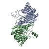 8hdnC 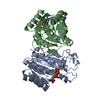 8hs4C 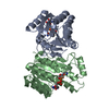 8hs5C 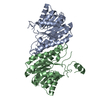 8hs6C 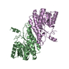 8hs9C 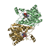 8hsaC C: citing same article ( |
|---|---|
| Similar structure data |
-Links
 PDBj
PDBj


- Assembly
| Deposited unit | 
| ||||||||
|---|---|---|---|---|---|---|---|---|---|
| 1 | 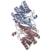
| ||||||||
| 2 | 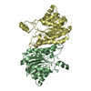
| ||||||||
| Unit cell |
|
-Components
| #1: Protein | Mass: 25854.598 Da / Num. of mol.: 4 / Fragment: 1-53 truncation / Mutation: K262T Source method: isolated from a genetically manipulated source Details: Sequence reference for WP_014489477 is not available in UniProt at the time of biocuration. Current sequence reference is from UniProt id Q8YIN7. Source: (gene. exp.) Brucella melitensis biotype 1 (strain 16M / ATCC 23456 / NCTC 10094) (bacteria)Gene: BMEI0406 / Production host: References: UniProt: Q8YIN7, 7alpha-hydroxysteroid dehydrogenase #2: Water | ChemComp-HOH / |  Mass: 18.015 Da / Num. of mol.: 736 / Source method: isolated from a natural source / Formula: H2O Mass: 18.015 Da / Num. of mol.: 736 / Source method: isolated from a natural source / Formula: H2OHas ligand of interest | Y | |
|---|
-Experimental details
-Experiment
| Experiment | Method: X-RAY DIFFRACTION / Number of used crystals: 1 |
|---|
- Sample preparation
| Crystal | Density Matthews: 2.58 Å3/Da / Density % sol: 52.39 % |
|---|---|
| Crystal grow | Temperature: 289 K / Method: vapor diffusion, sitting drop / pH: 4 / Details: 0.1M MIB, 10% PEG3350 / PH range: 4.0-8.0 |
-Data collection
| Diffraction | Mean temperature: 100 K / Serial crystal experiment: N |
|---|---|
| Diffraction source | Source: SYNCHROTRON / Site: SSRF / Beamline: BL17U / Wavelength: 1 Å |
| Detector | Type: DECTRIS EIGER X 16M / Detector: PIXEL / Date: Jul 12, 2021 |
| Radiation | Protocol: SINGLE WAVELENGTH / Monochromatic (M) / Laue (L): M / Scattering type: x-ray |
| Radiation wavelength | Wavelength: 1 Å / Relative weight: 1 |
| Reflection | Resolution: 1.99→43.18 Å / Num. obs: 73068 / % possible obs: 99.9 % / Redundancy: 9.9 % / Biso Wilson estimate: 23.48 Å2 / CC1/2: 0.996 / Net I/σ(I): 15.5 |
| Reflection shell | Resolution: 1.99→2.07 Å / Redundancy: 10.1 % / Mean I/σ(I) obs: 6.6 / Num. unique obs: 73517 / CC1/2: 0.972 / % possible all: 100 |
- Processing
| Software |
| ||||||||||||||||||||||||||||||||||||||||||||||||||||||||||||||||||||||||||||||||||||||||||||||||||||||||||||||||||||||||||||||||||||||||||||||||||||||||||||||||||||||||||||||||||||||
|---|---|---|---|---|---|---|---|---|---|---|---|---|---|---|---|---|---|---|---|---|---|---|---|---|---|---|---|---|---|---|---|---|---|---|---|---|---|---|---|---|---|---|---|---|---|---|---|---|---|---|---|---|---|---|---|---|---|---|---|---|---|---|---|---|---|---|---|---|---|---|---|---|---|---|---|---|---|---|---|---|---|---|---|---|---|---|---|---|---|---|---|---|---|---|---|---|---|---|---|---|---|---|---|---|---|---|---|---|---|---|---|---|---|---|---|---|---|---|---|---|---|---|---|---|---|---|---|---|---|---|---|---|---|---|---|---|---|---|---|---|---|---|---|---|---|---|---|---|---|---|---|---|---|---|---|---|---|---|---|---|---|---|---|---|---|---|---|---|---|---|---|---|---|---|---|---|---|---|---|---|---|---|---|
| Refinement | Method to determine structure: MOLECULAR REPLACEMENT / Resolution: 1.99→42.06 Å / SU ML: 0.16 / Cross valid method: FREE R-VALUE / σ(F): 1.96 / Phase error: 20.82 / Stereochemistry target values: ML
| ||||||||||||||||||||||||||||||||||||||||||||||||||||||||||||||||||||||||||||||||||||||||||||||||||||||||||||||||||||||||||||||||||||||||||||||||||||||||||||||||||||||||||||||||||||||
| Solvent computation | Shrinkage radii: 0.9 Å / VDW probe radii: 1.1 Å / Solvent model: FLAT BULK SOLVENT MODEL | ||||||||||||||||||||||||||||||||||||||||||||||||||||||||||||||||||||||||||||||||||||||||||||||||||||||||||||||||||||||||||||||||||||||||||||||||||||||||||||||||||||||||||||||||||||||
| Refinement step | Cycle: LAST / Resolution: 1.99→42.06 Å
| ||||||||||||||||||||||||||||||||||||||||||||||||||||||||||||||||||||||||||||||||||||||||||||||||||||||||||||||||||||||||||||||||||||||||||||||||||||||||||||||||||||||||||||||||||||||
| Refine LS restraints |
| ||||||||||||||||||||||||||||||||||||||||||||||||||||||||||||||||||||||||||||||||||||||||||||||||||||||||||||||||||||||||||||||||||||||||||||||||||||||||||||||||||||||||||||||||||||||
| LS refinement shell |
|
