 Movie
Movie Controller

[English] 日本語
 Yorodumi
- PDB-8hs4: Brucella melitensis 7-alpha-Hydroxysteroid Dehydrogenase mutant: ... -
+ Open data

- Basic information
| Entry | Database: PDB / ID: 8hs4 | |||||||||
|---|---|---|---|---|---|---|---|---|---|---|
| Title | Brucella melitensis 7-alpha-Hydroxysteroid Dehydrogenase mutant: 1-53 truncation/K262T-NAD+ | |||||||||
 Components Components | 7-alpha-hydroxysteroid dehydrogenase | |||||||||
 Keywords Keywords | OXIDOREDUCTASE / SDR family / hydroxysteroid dehydrogenase | |||||||||
| Function / homology |  Function and homology information Function and homology information7alpha-hydroxysteroid dehydrogenase / cholate 7-alpha-dehydrogenase (NAD+) activity / monocarboxylic acid metabolic process / lipid metabolic process / nucleotide binding Similarity search - Function | |||||||||
| Biological species |  Brucella melitensis biotype 1 (bacteria) Brucella melitensis biotype 1 (bacteria) | |||||||||
| Method |  X-RAY DIFFRACTION / SYNCHROTRON / MOLECULAR REPLACEMENT / Resolution: 2 Å X-RAY DIFFRACTION / SYNCHROTRON / MOLECULAR REPLACEMENT / Resolution: 2 Å | |||||||||
 Authors Authors | Liu, Z.Y. / Zhang, R.Z. | |||||||||
| Funding support |  China, 2items China, 2items
| |||||||||
 Citation Citation | Journal: To Be Published Title: Structure of 7-alpha-hydroxysteroid dehydrogenase Authors: Liu, Z.Y. / Zhang, R.Z. | |||||||||
| History |
|
- Structure visualization
| Structure viewer | Molecule: MolmilJmol/JSmol |
|---|
- Downloads & links
-Download
| PDBx/mmCIF format | 8hs4.cif.gz | 102.1 KB | Display | PDBx/mmCIF format |
|---|---|---|---|---|
| PDB format | pdb8hs4.ent.gz | 77 KB | Display | PDB format |
| PDBx/mmJSON format | 8hs4.json.gz | Tree view | PDBx/mmJSON format | |
| Others |  Other downloads Other downloads |
-Validation report
| Arichive directory | https://data.pdbj.org/pub/pdb/validation_reports/hs/8hs4ftp://data.pdbj.org/pub/pdb/validation_reports/hs/8hs4 | HTTPS FTP |
|---|
-Related structure data
| Related structure data |  8hdeC  8hdiC  8hdnC  8hs5C  8hs6C 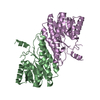 8hs9C 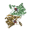 8hsaC C: citing same article ( |
|---|---|
| Similar structure data |
-Links
 PDBj
PDBj


- Assembly
| Deposited unit | 
| ||||||||
|---|---|---|---|---|---|---|---|---|---|
| 1 |
| ||||||||
| Unit cell |
|
-Components
| #1: Protein | Mass: 25495.154 Da / Num. of mol.: 2 / Mutation: K262T Source method: isolated from a genetically manipulated source Details: Author stated: in our study, wild-type 7alpha-hydroxysteroid dehydrogenase (protein_id="QEX87556.1") from Brucella melis strain RM57(RM57CP044342.1) was cloned into E. coli. Sequence ...Details: Author stated: in our study, wild-type 7alpha-hydroxysteroid dehydrogenase (protein_id="QEX87556.1") from Brucella melis strain RM57(RM57CP044342.1) was cloned into E. coli. Sequence reference for strain RM57 is not available in UniProt at the time of biocuration. Current sequence reference is from UniProt id Q8YIN7. Source: (gene. exp.) Brucella melitensis biotype 1 (strain 16M / ATCC 23456 / NCTC 10094) (bacteria)Strain: RM57 / Gene: BMEI0406 / Production host: References: UniProt: Q8YIN7, 7alpha-hydroxysteroid dehydrogenase #2: Chemical |   Mass: 663.425 Da / Num. of mol.: 2 / Source method: obtained synthetically / Formula: C21H27N7O14P2 / Feature type: SUBJECT OF INVESTIGATION / Comment: NAD*YM Mass: 663.425 Da / Num. of mol.: 2 / Source method: obtained synthetically / Formula: C21H27N7O14P2 / Feature type: SUBJECT OF INVESTIGATION / Comment: NAD*YM#3: Water | ChemComp-HOH / |  Mass: 18.015 Da / Num. of mol.: 145 / Source method: isolated from a natural source / Formula: H2O Mass: 18.015 Da / Num. of mol.: 145 / Source method: isolated from a natural source / Formula: H2OHas ligand of interest | Y | |
|---|
-Experimental details
-Experiment
| Experiment | Method: X-RAY DIFFRACTION / Number of used crystals: 1 |
|---|
- Sample preparation
| Crystal | Density Matthews: 2.52 Å3/Da / Density % sol: 51.23 % |
|---|---|
| Crystal grow | Temperature: 289 K / Method: vapor diffusion, sitting drop / pH: 4 / Details: 0.1M MIB, 10% PEG3350 / PH range: 4.0-8.0 |
-Data collection
| Diffraction | Mean temperature: 100 K / Serial crystal experiment: N |
|---|---|
| Diffraction source | Source: SYNCHROTRON / Site: SSRF / Beamline: BL17U / Wavelength: 0.9875 Å |
| Detector | Type: DECTRIS EIGER X 16M / Detector: PIXEL / Date: Jul 12, 2021 |
| Radiation | Protocol: SINGLE WAVELENGTH / Monochromatic (M) / Laue (L): M / Scattering type: x-ray |
| Radiation wavelength | Wavelength: 0.9875 Å / Relative weight: 1 |
| Reflection | Resolution: 2→40.88 Å / Num. obs: 72757 / % possible obs: 99.9 % / Redundancy: 9.9 % / CC1/2: 0.996 / Rmerge(I) obs: 0.112 / Rrim(I) all: 0.118 / Net I/σ(I): 15.5 |
| Reflection shell | Resolution: 2→2.073 Å / Num. unique obs: 72757 / CC1/2: 0.973 |
- Processing
| Software |
| |||||||||||||||||||||||||||||||||||||||||||||||||||||||||||||||||||||||||||||||||||||||||||||||||||||||||
|---|---|---|---|---|---|---|---|---|---|---|---|---|---|---|---|---|---|---|---|---|---|---|---|---|---|---|---|---|---|---|---|---|---|---|---|---|---|---|---|---|---|---|---|---|---|---|---|---|---|---|---|---|---|---|---|---|---|---|---|---|---|---|---|---|---|---|---|---|---|---|---|---|---|---|---|---|---|---|---|---|---|---|---|---|---|---|---|---|---|---|---|---|---|---|---|---|---|---|---|---|---|---|---|---|---|---|
| Refinement | Method to determine structure: MOLECULAR REPLACEMENT / Resolution: 2→40.85 Å / SU ML: 0.17 / Cross valid method: THROUGHOUT / σ(F): 1.36 / Phase error: 19.59 / Stereochemistry target values: ML
| |||||||||||||||||||||||||||||||||||||||||||||||||||||||||||||||||||||||||||||||||||||||||||||||||||||||||
| Solvent computation | Shrinkage radii: 0.9 Å / VDW probe radii: 1.1 Å / Solvent model: FLAT BULK SOLVENT MODEL | |||||||||||||||||||||||||||||||||||||||||||||||||||||||||||||||||||||||||||||||||||||||||||||||||||||||||
| Refinement step | Cycle: LAST / Resolution: 2→40.85 Å
| |||||||||||||||||||||||||||||||||||||||||||||||||||||||||||||||||||||||||||||||||||||||||||||||||||||||||
| Refine LS restraints | Type: f_plane_restr / Dev ideal: 0.01 / Number: 626 | |||||||||||||||||||||||||||||||||||||||||||||||||||||||||||||||||||||||||||||||||||||||||||||||||||||||||
| LS refinement shell |
|
