 Movie
Movie Controller

[English] 日本語
 Yorodumi
- PDB-8h62: Crystal structure of Internalin A from Listeria monocytogenes wit... -
+ Open data

- Basic information
| Entry | Database: PDB / ID: 8h62 | ||||||
|---|---|---|---|---|---|---|---|
| Title | Crystal structure of Internalin A from Listeria monocytogenes with human E-cadherin EC12 | ||||||
 Components Components |
| ||||||
 Keywords Keywords | CELL INVASION / Internalin A Cadherin Bacterial invasion nanobody Surface plasmon resonance Isothermal titration calorimetry | ||||||
| Function / homology |  Function and homology information Function and homology informationresponse to heparin / desmosome assembly / response to Gram-positive bacterium / pituitary gland development / gamma-catenin binding / Regulation of MITF-M-dependent genes involved in extracellular matrix, focal adhesion and epithelial-to-mesenchymal transition / negative regulation of axon extension / desmosome / cellular response to indole-3-methanol / calcium-dependent cell-cell adhesion ...response to heparin / desmosome assembly / response to Gram-positive bacterium / pituitary gland development / gamma-catenin binding / Regulation of MITF-M-dependent genes involved in extracellular matrix, focal adhesion and epithelial-to-mesenchymal transition / negative regulation of axon extension / desmosome / cellular response to indole-3-methanol / calcium-dependent cell-cell adhesion / flotillin complex / regulation of protein catabolic process at postsynapse, modulating synaptic transmission / cell-cell adhesion mediated by cadherin / adherens junction organization / Formation of definitive endoderm / catenin complex / Apoptotic cleavage of cell adhesion proteins / cell-cell junction assembly / Adherens junctions interactions / GTPase activating protein binding / ankyrin binding / negative regulation of cell-cell adhesion / cellular response to lithium ion / apical junction complex / homophilic cell-cell adhesion / lateral plasma membrane / Integrin cell surface interactions / RHO GTPases activate IQGAPs / positive regulation of protein localization / Degradation of the extracellular matrix / synapse assembly / cell adhesion molecule binding / Transcriptional and post-translational regulation of MITF-M expression and activity / InlA-mediated entry of Listeria monocytogenes into host cells / peptidoglycan-based cell wall / protein tyrosine kinase binding / negative regulation of cell migration / protein localization to plasma membrane / adherens junction / trans-Golgi network / cell-cell adhesion / beta-catenin binding / positive regulation of protein import into nucleus / response to toxic substance / cytoplasmic side of plasma membrane / cell morphogenesis / neuron projection development / Immunoregulatory interactions between a Lymphoid and a non-Lymphoid cell / cell junction / cell migration / actin cytoskeleton / lamellipodium / regulation of gene expression / endosome / postsynapse / cadherin binding / response to xenobiotic stimulus / calcium ion binding / positive regulation of DNA-templated transcription / perinuclear region of cytoplasm / glutamatergic synapse / extracellular exosome / extracellular region / identical protein binding / membrane / plasma membrane / cytoplasm Similarity search - Function | ||||||
| Biological species |  Listeria monocytogenes serovar 1/2a (bacteria) Listeria monocytogenes serovar 1/2a (bacteria) Homo sapiens (human) Homo sapiens (human) | ||||||
| Method |  X-RAY DIFFRACTION / SYNCHROTRON / MOLECULAR REPLACEMENT / Resolution: 1.91 Å X-RAY DIFFRACTION / SYNCHROTRON / MOLECULAR REPLACEMENT / Resolution: 1.91 Å | ||||||
 Authors Authors | Caaveiro, J.M.M. / Nagatoish, S. / Tsumoto, K. | ||||||
| Funding support |  Japan, 1items Japan, 1items
| ||||||
 Citation Citation | Journal: J.Biol.Chem. / Year: 2023 Title: Anti-InlA single-domain antibodies that inhibit the cell invasion of Listeria monocytogenes. Authors: Yamazaki, T. / Nagatoishi, S. / Yamawaki, T. / Nozawa, T. / Matsunaga, R. / Nakakido, M. / Caaveiro, J.M.M. / Nakagawa, I. / Tsumoto, K. | ||||||
| History |
|
- Structure visualization
| Structure viewer | Molecule: MolmilJmol/JSmol |
|---|
- Downloads & links
-Download
| PDBx/mmCIF format | 8h62.cif.gz | 284 KB | Display | PDBx/mmCIF format |
|---|---|---|---|---|
| PDB format | pdb8h62.ent.gz | 225.4 KB | Display | PDB format |
| PDBx/mmJSON format | 8h62.json.gz | Tree view | PDBx/mmJSON format | |
| Others |  Other downloads Other downloads |
-Validation report
| Arichive directory | https://data.pdbj.org/pub/pdb/validation_reports/h6/8h62ftp://data.pdbj.org/pub/pdb/validation_reports/h6/8h62 | HTTPS FTP |
|---|
-Related structure data
| Related structure data |  8h63C 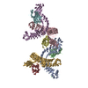 8h64C  1o6sS  4zmtS S: Starting model for refinement C: citing same article ( |
|---|---|
| Similar structure data |
-Links
 PDBj
PDBj


- Assembly
| Deposited unit | 
| ||||||||
|---|---|---|---|---|---|---|---|---|---|
| 1 |
| ||||||||
| Unit cell |
|
-Components
| #1: Protein | Mass: 49914.758 Da / Num. of mol.: 1 Source method: isolated from a genetically manipulated source Source: (gene. exp.) Listeria monocytogenes serovar 1/2a (bacteria)Strain: ATCC BAA-679 / EGD-e / Gene: inlA, lmo0433, intlA / Production host: | ||||||
|---|---|---|---|---|---|---|---|
| #2: Protein | Mass: 23134.660 Da / Num. of mol.: 1 / Mutation: C163S Source method: isolated from a genetically manipulated source Source: (gene. exp.) Homo sapiens (human) / Gene: CDH1, CDHE, UVO / Production host: | ||||||
| #3: Chemical | ChemComp-CA /   Mass: 40.078 Da / Num. of mol.: 4 / Source method: obtained synthetically / Formula: Ca / Feature type: SUBJECT OF INVESTIGATION Mass: 40.078 Da / Num. of mol.: 4 / Source method: obtained synthetically / Formula: Ca / Feature type: SUBJECT OF INVESTIGATION#4: Chemical | ChemComp-ACT / |   Mass: 59.044 Da / Num. of mol.: 1 / Source method: obtained synthetically / Formula: C2H3O2 Mass: 59.044 Da / Num. of mol.: 1 / Source method: obtained synthetically / Formula: C2H3O2#5: Water | ChemComp-HOH / |  Mass: 18.015 Da / Num. of mol.: 630 / Source method: isolated from a natural source / Formula: H2O Mass: 18.015 Da / Num. of mol.: 630 / Source method: isolated from a natural source / Formula: H2OHas ligand of interest | Y | |
-Experimental details
-Experiment
| Experiment | Method: X-RAY DIFFRACTION / Number of used crystals: 1 |
|---|
- Sample preparation
| Crystal | Density Matthews: 3.53 Å3/Da / Density % sol: 65.12 % |
|---|---|
| Crystal grow | Temperature: 293.15 K / Method: vapor diffusion, sitting drop / Details: 0.2 M Ammonium acetate 45% MPD |
-Data collection
| Diffraction | Mean temperature: 100 K / Serial crystal experiment: N |
|---|---|
| Diffraction source | Source: SYNCHROTRON / Site: Photon Factory / Beamline: BL-5A / Wavelength: 1 Å |
| Detector | Type: ADSC QUANTUM 315 / Detector: CCD / Date: May 12, 2017 |
| Radiation | Protocol: SINGLE WAVELENGTH / Monochromatic (M) / Laue (L): M / Scattering type: x-ray |
| Radiation wavelength | Wavelength: 1 Å / Relative weight: 1 |
| Reflection | Resolution: 1.91→34.8 Å / Num. obs: 71483 / % possible obs: 92.3 % / Redundancy: 3.8 % / CC1/2: 0.996 / Rmerge(I) obs: 0.088 / Net I/σ(I): 8.8 |
| Reflection shell | Resolution: 1.91→1.95 Å / Redundancy: 3.7 % / Rmerge(I) obs: 0.76 / Mean I/σ(I) obs: 2.1 / Num. unique obs: 4418 / CC1/2: 0.837 / % possible all: 92.1 |
- Processing
| Software |
| |||||||||||||||||||||||||||||||||||||||||||||||||||||||||||||||||||||||||||
|---|---|---|---|---|---|---|---|---|---|---|---|---|---|---|---|---|---|---|---|---|---|---|---|---|---|---|---|---|---|---|---|---|---|---|---|---|---|---|---|---|---|---|---|---|---|---|---|---|---|---|---|---|---|---|---|---|---|---|---|---|---|---|---|---|---|---|---|---|---|---|---|---|---|---|---|---|
| Refinement | Method to determine structure: MOLECULAR REPLACEMENT Starting model: 1o6s 4zmt Resolution: 1.91→34.8 Å / Cor.coef. Fo:Fc: 0.961 / Cor.coef. Fo:Fc free: 0.945 / SU B: 6.527 / SU ML: 0.097 / Cross valid method: THROUGHOUT / σ(F): 0 / ESU R: 0.125 / ESU R Free: 0.121 / Stereochemistry target values: MAXIMUM LIKELIHOOD Details: HYDROGENS HAVE BEEN ADDED IN THE RIDING POSITIONS U VALUES : WITH TLS ADDED
| |||||||||||||||||||||||||||||||||||||||||||||||||||||||||||||||||||||||||||
| Solvent computation | Ion probe radii: 0.8 Å / Shrinkage radii: 0.8 Å / VDW probe radii: 1.2 Å / Solvent model: MASK | |||||||||||||||||||||||||||||||||||||||||||||||||||||||||||||||||||||||||||
| Displacement parameters | Biso max: 92.26 Å2 / Biso mean: 26.343 Å2 / Biso min: 14.13 Å2
| |||||||||||||||||||||||||||||||||||||||||||||||||||||||||||||||||||||||||||
| Refinement step | Cycle: final / Resolution: 1.91→34.8 Å
| |||||||||||||||||||||||||||||||||||||||||||||||||||||||||||||||||||||||||||
| Refine LS restraints |
| |||||||||||||||||||||||||||||||||||||||||||||||||||||||||||||||||||||||||||
| LS refinement shell | Resolution: 1.91→1.959 Å / Rfactor Rfree error: 0 / Total num. of bins used: 20
| |||||||||||||||||||||||||||||||||||||||||||||||||||||||||||||||||||||||||||
| Refinement TLS params. | Method: refined / Refine-ID: X-RAY DIFFRACTION
| |||||||||||||||||||||||||||||||||||||||||||||||||||||||||||||||||||||||||||
| Refinement TLS group |
|
