 Movie
Movie Controller


+ Open data

- Basic information
| Entry | Database: PDB / ID: 8g89 | ||||||
|---|---|---|---|---|---|---|---|
| Title | HSD17B13 in complex with cofactor and inhibitor | ||||||
 Components Components | Hydroxysteroid 17-beta dehydrogenase 13 | ||||||
 Keywords Keywords | OXIDOREDUCTASE/INHIBITOR / 17 beta-hydroxysteroid dehydrogenase 13 / lipid droplet associated protein / inhibitor / OXIDOREDUCTASE / OXIDOREDUCTASE-INHIBITOR complex | ||||||
| Function / homology |  Function and homology information Function and homology information17beta-estradiol 17-dehydrogenase / estradiol 17-beta-dehydrogenase [NAD(P)+] activity / lipid droplet / lipid metabolic process / nucleotide binding / endoplasmic reticulum Similarity search - Function | ||||||
| Biological species |  | ||||||
| Method |  X-RAY DIFFRACTION / SYNCHROTRON / FOURIER SYNTHESIS / Resolution: 2.222 Å X-RAY DIFFRACTION / SYNCHROTRON / FOURIER SYNTHESIS / Resolution: 2.222 Å | ||||||
 Authors Authors | Liu, S. | ||||||
| Funding support |  United States, 1items United States, 1items
| ||||||
 Citation Citation | Journal: Nat Commun / Year: 2023 Title: Structural basis of lipid-droplet localization of 17-beta-hydroxysteroid dehydrogenase 13. Authors: Liu, S. / Sommese, R.F. / Nedoma, N.L. / Stevens, L.M. / Dutra, J.K. / Zhang, L. / Edmonds, D.J. / Wang, Y. / Garnsey, M. / Clasquin, M.F. | ||||||
| History |
|
- Structure visualization
| Structure viewer | Molecule: MolmilJmol/JSmol |
|---|
- Downloads & links
-Download
| PDBx/mmCIF format | 8g89.cif.gz | 245.8 KB | Display | PDBx/mmCIF format |
|---|---|---|---|---|
| PDB format | pdb8g89.ent.gz | 200.2 KB | Display | PDB format |
| PDBx/mmJSON format | 8g89.json.gz | Tree view | PDBx/mmJSON format | |
| Others |  Other downloads Other downloads |
-Validation report
| Arichive directory | https://data.pdbj.org/pub/pdb/validation_reports/g8/8g89ftp://data.pdbj.org/pub/pdb/validation_reports/g8/8g89 | HTTPS FTP |
|---|
-Related structure data
| Related structure data | 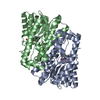 8g84SC 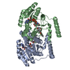 8g93C 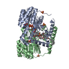 8g9vC S: Starting model for refinement C: citing same article ( |
|---|---|
| Similar structure data |
-Links
 PDBj
PDBj


- Assembly
| Deposited unit | 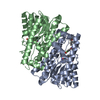
| ||||||||
|---|---|---|---|---|---|---|---|---|---|
| 1 |
| ||||||||
| Unit cell |
|
-Components
| #1: Protein | Mass: 35364.211 Da / Num. of mol.: 2 Source method: isolated from a genetically manipulated source Source: (gene. exp.)   Spodoptera frugiperda (fall armyworm) / References: UniProt: A0A8C0PP93 Spodoptera frugiperda (fall armyworm) / References: UniProt: A0A8C0PP93#2: Chemical |   Mass: 663.425 Da / Num. of mol.: 2 / Source method: obtained synthetically / Formula: C21H27N7O14P2 / Feature type: SUBJECT OF INVESTIGATION / Comment: NAD*YM Mass: 663.425 Da / Num. of mol.: 2 / Source method: obtained synthetically / Formula: C21H27N7O14P2 / Feature type: SUBJECT OF INVESTIGATION / Comment: NAD*YM#3: Chemical | ChemComp-YXW / |   Mass: 391.408 Da / Num. of mol.: 1 / Source method: obtained synthetically / Formula: C21H23F2NO4 / Feature type: SUBJECT OF INVESTIGATION Mass: 391.408 Da / Num. of mol.: 1 / Source method: obtained synthetically / Formula: C21H23F2NO4 / Feature type: SUBJECT OF INVESTIGATION#4: Water | ChemComp-HOH / |  Mass: 18.015 Da / Num. of mol.: 81 / Source method: isolated from a natural source / Formula: H2O Mass: 18.015 Da / Num. of mol.: 81 / Source method: isolated from a natural source / Formula: H2OHas ligand of interest | Y | |
|---|
-Experimental details
-Experiment
| Experiment | Method: X-RAY DIFFRACTION / Number of used crystals: 1 |
|---|
- Sample preparation
| Crystal | Density Matthews: 3.32 Å3/Da / Density % sol: 62.99 % |
|---|---|
| Crystal grow | Temperature: 293 K / Method: vapor diffusion, sitting drop / Details: 30% PEG3350, 0.2 M ammonium chloride |
-Data collection
| Diffraction | Mean temperature: 100 K / Serial crystal experiment: N |
|---|---|
| Diffraction source | Source: SYNCHROTRON / Site: APS / Beamline: 17-ID / Wavelength: 1 Å |
| Detector | Type: DECTRIS EIGER X 16M / Detector: PIXEL / Date: Oct 2, 2019 |
| Radiation | Protocol: SINGLE WAVELENGTH / Monochromatic (M) / Laue (L): M / Scattering type: x-ray |
| Radiation wavelength | Wavelength: 1 Å / Relative weight: 1 |
| Reflection | Resolution: 2.23→71.19 Å / Num. obs: 32599 / % possible obs: 92.9 % / Redundancy: 6.6 % / CC1/2: 0.999 / Rpim(I) all: 0.039 / Net I/σ(I): 14.2 |
| Reflection shell | Resolution: 2.23→2.47 Å / Num. unique obs: 1631 / CC1/2: 0.79 / Rpim(I) all: 0.57 |
- Processing
| Software |
| |||||||||||||||||||||||||||||||||||||||||||||||||||||||||||||||||||||||||||
|---|---|---|---|---|---|---|---|---|---|---|---|---|---|---|---|---|---|---|---|---|---|---|---|---|---|---|---|---|---|---|---|---|---|---|---|---|---|---|---|---|---|---|---|---|---|---|---|---|---|---|---|---|---|---|---|---|---|---|---|---|---|---|---|---|---|---|---|---|---|---|---|---|---|---|---|---|
| Refinement | Method to determine structure: FOURIER SYNTHESIS Starting model: 8G84 Resolution: 2.222→23.16 Å / Cor.coef. Fo:Fc: 0.942 / Cor.coef. Fo:Fc free: 0.93 / SU R Cruickshank DPI: 0.311 / Cross valid method: THROUGHOUT / SU R Blow DPI: 0.304 / SU Rfree Blow DPI: 0.214 / SU Rfree Cruickshank DPI: 0.218
| |||||||||||||||||||||||||||||||||||||||||||||||||||||||||||||||||||||||||||
| Displacement parameters | Biso mean: 65.66 Å2
| |||||||||||||||||||||||||||||||||||||||||||||||||||||||||||||||||||||||||||
| Refine analyze | Luzzati coordinate error obs: 0.33 Å | |||||||||||||||||||||||||||||||||||||||||||||||||||||||||||||||||||||||||||
| Refinement step | Cycle: LAST / Resolution: 2.222→23.16 Å
| |||||||||||||||||||||||||||||||||||||||||||||||||||||||||||||||||||||||||||
| Refine LS restraints |
| |||||||||||||||||||||||||||||||||||||||||||||||||||||||||||||||||||||||||||
| LS refinement shell | Resolution: 2.222→2.39 Å
| |||||||||||||||||||||||||||||||||||||||||||||||||||||||||||||||||||||||||||
| Refinement TLS params. | Refine-ID: X-RAY DIFFRACTION
| |||||||||||||||||||||||||||||||||||||||||||||||||||||||||||||||||||||||||||
| Refinement TLS group |
|
