 Movie
Movie Controller

[English] 日本語
 Yorodumi
- PDB-8g1h: Ancestral protein AncTh of Phosphomethylpirimidine kinases family -
+ Open data

- Basic information
| Entry | Database: PDB / ID: 8g1h | |||||||||
|---|---|---|---|---|---|---|---|---|---|---|
| Title | Ancestral protein AncTh of Phosphomethylpirimidine kinases family | |||||||||
 Components Components | Phosphomethylpyrimidine Kinase | |||||||||
 Keywords Keywords | TRANSFERASE / Ancestral protein reconstruction / 5-phosphohydroxymethyl-2-methylpirimidine kinase / phosphomethylpirimidine kinases family / enzyme evolution | |||||||||
| Function / homology | PHOSPHATE ION Function and homology information Function and homology information | |||||||||
| Biological species | synthetic construct (others) | |||||||||
| Method |  X-RAY DIFFRACTION / SYNCHROTRON / MOLECULAR REPLACEMENT / Resolution: 2.7 Å X-RAY DIFFRACTION / SYNCHROTRON / MOLECULAR REPLACEMENT / Resolution: 2.7 Å | |||||||||
 Authors Authors | Perez, M. / Munoz, S. / Cea, P. / Castro-Fernandez, V. | |||||||||
| Funding support |  Chile, 2items Chile, 2items
| |||||||||
 Citation Citation | Journal: Mol.Biol.Evol. / Year: 2024 Title: Deciphering Structural Traits for Thermal and Kinetic Stability across Protein Family Evolution through Ancestral Sequence Reconstruction. Authors: Cea, P.A. / Perez, M. / Herrera, S.M. / Munoz, S.M. / Fuentes-Ugarte, N. / Coche-Miranda, J. / Maturana, P. / Guixe, V. / Castro-Fernandez, V. | |||||||||
| History |
|
- Structure visualization
| Structure viewer | Molecule: MolmilJmol/JSmol |
|---|
- Downloads & links
-Download
| PDBx/mmCIF format | 8g1h.cif.gz | 66.3 KB | Display | PDBx/mmCIF format |
|---|---|---|---|---|
| PDB format | pdb8g1h.ent.gz | 43 KB | Display | PDB format |
| PDBx/mmJSON format | 8g1h.json.gz | Tree view | PDBx/mmJSON format | |
| Others |  Other downloads Other downloads |
-Validation report
| Summary document | 8g1h_validation.pdf.gz | 435.6 KB | Display | wwPDB validaton report |
|---|---|---|---|---|
| Full document | 8g1h_full_validation.pdf.gz | 436.5 KB | Display | |
| Data in XML | 8g1h_validation.xml.gz | 11.1 KB | Display | |
| Data in CIF | 8g1h_validation.cif.gz | 14.2 KB | Display | |
| Arichive directory | https://data.pdbj.org/pub/pdb/validation_reports/g1/8g1hftp://data.pdbj.org/pub/pdb/validation_reports/g1/8g1h | HTTPS FTP |
-Related structure data
| Related structure data | 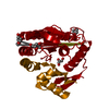 1ub0S S: Starting model for refinement |
|---|---|
| Similar structure data |


-Links
 PDBj
PDBj- Assembly
| Deposited unit | 
| ||||||||||||
|---|---|---|---|---|---|---|---|---|---|---|---|---|---|
| 1 | 
| ||||||||||||
| Unit cell |
|
-Components
| #1: Protein | Mass: 26798.918 Da / Num. of mol.: 1 Source method: isolated from a genetically manipulated source Source: (gene. exp.) synthetic construct (others) / Production host:  | ||||||||
|---|---|---|---|---|---|---|---|---|---|
| #2: Chemical |   Mass: 62.068 Da / Num. of mol.: 2 / Source method: obtained synthetically / Formula: C2H6O2 Mass: 62.068 Da / Num. of mol.: 2 / Source method: obtained synthetically / Formula: C2H6O2#3: Chemical |   Mass: 94.971 Da / Num. of mol.: 2 / Source method: obtained synthetically / Formula: PO4 Mass: 94.971 Da / Num. of mol.: 2 / Source method: obtained synthetically / Formula: PO4#4: Water | ChemComp-HOH / |  Mass: 18.015 Da / Num. of mol.: 27 / Source method: isolated from a natural source / Formula: H2O Mass: 18.015 Da / Num. of mol.: 27 / Source method: isolated from a natural source / Formula: H2OHas ligand of interest | N | Has protein modification | Y | |
-Experimental details
-Experiment
| Experiment | Method: X-RAY DIFFRACTION / Number of used crystals: 1 |
|---|
- Sample preparation
| Crystal | Density Matthews: 2.43 Å3/Da / Density % sol: 49.45 % |
|---|---|
| Crystal grow | Temperature: 292 K / Method: vapor diffusion, sitting drop / pH: 7.5 Details: Protein conditions: Protein 26 mg/ml, Tris-HCl 25 mM, NaCl 0.3 M, b-Mercaptoethanol 5 mM, ATP 1 mM, MgCl2 5 mM. Reservoir condition: Ethylene glycols 0.12 M (0.3M Diethylene glycol; 0.3M ...Details: Protein conditions: Protein 26 mg/ml, Tris-HCl 25 mM, NaCl 0.3 M, b-Mercaptoethanol 5 mM, ATP 1 mM, MgCl2 5 mM. Reservoir condition: Ethylene glycols 0.12 M (0.3M Diethylene glycol; 0.3M Triethylene glycol; 0.3M Tetraethylene glycol; 0.3M Pentaethylene glycol), Sodium HEPES/ MOPS pH 7.5, 37.5% precipitants (MPD, PEG 1000, PEG 3350) |
-Data collection
| Diffraction | Mean temperature: 100 K / Serial crystal experiment: N |
|---|---|
| Diffraction source | Source: SYNCHROTRON / Site: LNLS SIRIUS / Beamline: MANACA / Wavelength: 0.97718 Å |
| Detector | Type: DECTRIS PILATUS 2M / Detector: PIXEL / Date: Oct 7, 2022 |
| Radiation | Monochromator: Double crystal Monochromator - water- cooled Si(111) Protocol: SINGLE WAVELENGTH / Monochromatic (M) / Laue (L): M / Scattering type: x-ray |
| Radiation wavelength | Wavelength: 0.97718 Å / Relative weight: 1 |
| Reflection | Resolution: 2.7→44.01 Å / Num. obs: 8030 / % possible obs: 99.84 % / Redundancy: 34.1 % / Biso Wilson estimate: 54.5 Å2 / CC1/2: 0.999 / CC star: 1 / Rmerge(I) obs: 0.1926 / Rpim(I) all: 0.033 / Rrim(I) all: 0.1956 / Net I/av σ(I): 21.62 / Net I/σ(I): 21.62 |
| Reflection shell | Resolution: 2.7→2.797 Å / Rmerge(I) obs: 1.275 / Mean I/σ(I) obs: 4.05 / Num. unique obs: 753 / CC1/2: 0.94 / CC star: 0.984 / Rpim(I) all: 0.2123 / Rrim(I) all: 1.293 / % possible all: 99.87 |
- Processing
| Software |
| ||||||||||||||||||||||||||||
|---|---|---|---|---|---|---|---|---|---|---|---|---|---|---|---|---|---|---|---|---|---|---|---|---|---|---|---|---|---|
| Refinement | Method to determine structure: MOLECULAR REPLACEMENT Starting model: 1UB0 Resolution: 2.7→44.01 Å / SU ML: 0.3315 / Cross valid method: FREE R-VALUE / σ(F): 1.34 / Phase error: 21.025 Stereochemistry target values: GeoStd + Monomer Library + CDL v1.2
| ||||||||||||||||||||||||||||
| Solvent computation | Shrinkage radii: 0.9 Å / VDW probe radii: 1.11 Å / Solvent model: FLAT BULK SOLVENT MODEL | ||||||||||||||||||||||||||||
| Displacement parameters | Biso mean: 48.01 Å2 | ||||||||||||||||||||||||||||
| Refinement step | Cycle: LAST / Resolution: 2.7→44.01 Å
| ||||||||||||||||||||||||||||
| Refine LS restraints |
| ||||||||||||||||||||||||||||
| LS refinement shell |
|
