Resolution: 2.05→40 Å / Isotropic thermal model: Overall / Cross valid method: THROUGHOUT / σ(F): 0 / Stereochemistry target values: Engh & Huber Details: The electron density is poor for sidechain atoms of residues: LYS A117, LEU A139, ARG A189
Rfactor
Num. reflection
% reflection
Selection details
Rfree
0.2316
1046
-
RANDOM
Rwork
0.2066
-
-
-
all
-
22162
-
-
obs
-
21300
96.1 %
-
Displacement parameters
Biso mean: 43.3 Å2
Baniso -1
Baniso -2
Baniso -3
1-
3.74 Å2
2.42 Å2
0 Å2
2-
-
3.74 Å2
0 Å2
3-
-
-
-7.49 Å2
Refine analyze
Free
Obs
Luzzati coordinate error
0.28 Å
0.23 Å
Luzzati d res low
-
5 Å
Luzzati sigma a
0.2 Å
0.17 Å
Refinement step
Cycle: LAST / Resolution: 2.05→40 Å
Protein
Nucleic acid
Ligand
Solvent
Total
Num. atoms
1805
0
41
110
1956
Refine LS restraints
Refine-ID
Type
Dev ideal
X-RAY DIFFRACTION
c_bond_d
1.2
X-RAY DIFFRACTION
c_angle_deg
0.005
X-RAY DIFFRACTION
c_dihedral_angle_d
21.7
X-RAY DIFFRACTION
c_improper_angle_d
0.82
LS refinement shell
Refine-ID: X-RAY DIFFRACTION / Total num. of bins used: 6
Resolution (Å)
Rfactor Rfree
Num. reflection Rfree
Rfactor Rwork
Rfactor Rfree error
Num. reflection obs
% reflection obs (%)
2.05-2.14
0.266
116
0.235
0.025
2491
91.9
2.14-2.26
0.281
123
0.239
0.025
2530
93.4
2.26-2.4
0.23
117
0.225
0.021
2555
93.4
2.4-2.58
0.254
139
0.217
0.022
2606
95.7
2.58-2.84
0.235
143
0.2
0.02
2669
97.3
2.84-3.25
0.259
111
0.2
0.025
2712
98.4
+
About Yorodumi
-
News
-
Feb 9, 2022. New format data for meta-information of EMDB entries
New format data for meta-information of EMDB entries
Version 3 of the EMDB header file is now the official format.
The previous official version 1.9 will be removed from the archive.
In the structure databanks used in Yorodumi, some data are registered as the other names, "COVID-19 virus" and "2019-nCoV". Here are the details of the virus and the list of structure data.
Jan 31, 2019. EMDB accession codes are about to change! (news from PDBe EMDB page)
EMDB accession codes are about to change! (news from PDBe EMDB page)
The allocation of 4 digits for EMDB accession codes will soon come to an end. Whilst these codes will remain in use, new EMDB accession codes will include an additional digit and will expand incrementally as the available range of codes is exhausted. The current 4-digit format prefixed with “EMD-” (i.e. EMD-XXXX) will advance to a 5-digit format (i.e. EMD-XXXXX), and so on. It is currently estimated that the 4-digit codes will be depleted around Spring 2019, at which point the 5-digit format will come into force.
The EM Navigator/Yorodumi systems omit the EMD- prefix.
Related info.:Q: What is EMD? / ID/Accession-code notation in Yorodumi/EM Navigator
Yorodumi is a browser for structure data from EMDB, PDB, SASBDB, etc.
This page is also the successor to EM Navigator detail page, and also detail information page/front-end page for Omokage search.
The word "yorodu" (or yorozu) is an old Japanese word meaning "ten thousand". "mi" (miru) is to see.
Related info.:EMDB / PDB / SASBDB / Comparison of 3 databanks / Yorodumi Search / Aug 31, 2016. New EM Navigator & Yorodumi / Yorodumi Papers / Jmol/JSmol / Function and homology information / Changes in new EM Navigator and Yorodumi
 Movie
Movie Controller

 Yorodumi
 Open data
 Basic information
 Components
 Keywords
 Function and homology information
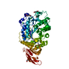
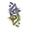
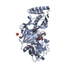
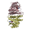
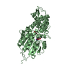
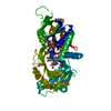
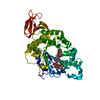
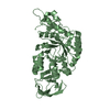
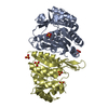
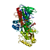
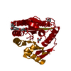
 Thermus thermophilus (bacteria)
 X-RAY DIFFRACTION /
  Authors
 Citation
 Structure visualization
 Downloads & links
 Other downloads
 PDBj
PDBj Assembly
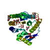

 Mass: 96.063 Da / Num. of mol.: 1 / Source method: obtained synthetically / Formula: SO4
 Mass: 88.105 Da / Num. of mol.: 2 / Source method: obtained synthetically / Formula: C4H8O2
 Mass: 92.094 Da / Num. of mol.: 4 / Source method: obtained synthetically / Formula: C3H8O3
 Mass: 18.015 Da / Num. of mol.: 110 / Source method: isolated from a natural source / Formula: H2O
 Sample preparation
 / Beamline: BL44B2 / Wavelength: 1 Å
 Processing