 Movie
Movie Controller

[English] 日本語
 Yorodumi
- PDB-2i5b: The crystal structure of an ADP complex of Bacillus subtilis pyri... -
+ Open data

- Basic information
| Entry | Database: PDB / ID: 2i5b | ||||||
|---|---|---|---|---|---|---|---|
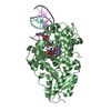
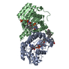
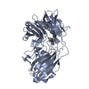
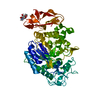
| Title | The crystal structure of an ADP complex of Bacillus subtilis pyridoxal kinase provides evidence for the parralel emergence of enzyme activity during evolution | ||||||
 Components Components | Phosphomethylpyrimidine kinase | ||||||
 Keywords Keywords | TRANSFERASE / ADP complex / PdxK / thiD / ribokinase superfamily | ||||||
| Function / homology |  Function and homology information Function and homology informationphosphomethylpyrimidine kinase activity / hydroxymethylpyrimidine kinase activity / pyridoxal kinase / pyridoxal kinase activity / thiamine biosynthetic process / ATP binding / metal ion binding / cytosol Similarity search - Function | ||||||
| Biological species |  | ||||||
| Method |  X-RAY DIFFRACTION / MOLECULAR REPLACEMENT / Resolution: 2.8 Å X-RAY DIFFRACTION / MOLECULAR REPLACEMENT / Resolution: 2.8 Å | ||||||
 Authors Authors | Newman, J.A. / Das, S.K. / Sedelnikova, S.E. / Rice, D.W. | ||||||
 Citation Citation | Journal: J.Mol.Biol. / Year: 2006 Title: The Crystal Structure of an ADP Complex of Bacillus subtilis Pyridoxal Kinase Provides Evidence for the Parallel Emergence of Enzyme Activity During Evolution. Authors: Newman, J.A. / Das, S.K. / Sedelnikova, S.E. / Rice, D.W. #1: Journal: To be publishedTitle: Cloning, purification and preliminary crystallographic analysis of a putative Pyridoxal Kinase from Bacillus subtilis | ||||||
| History |
|
- Structure visualization
| Structure viewer | Molecule: MolmilJmol/JSmol |
|---|
- Downloads & links
-Download
| PDBx/mmCIF format | 2i5b.cif.gz | 249.7 KB | Display | PDBx/mmCIF format |
|---|---|---|---|---|
| PDB format | pdb2i5b.ent.gz | 203.3 KB | Display | PDB format |
| PDBx/mmJSON format | 2i5b.json.gz | Tree view | PDBx/mmJSON format | |
| Others |  Other downloads Other downloads |
-Validation report
| Arichive directory | https://data.pdbj.org/pub/pdb/validation_reports/i5/2i5bftp://data.pdbj.org/pub/pdb/validation_reports/i5/2i5b | HTTPS FTP |
|---|
-Related structure data
| Related structure data | 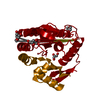 1ub0S S: Starting model for refinement |
|---|---|
| Similar structure data |


-Links
 PDBj
PDBj- Assembly
| Deposited unit | 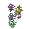
| |||||||||||||||||||||||||
|---|---|---|---|---|---|---|---|---|---|---|---|---|---|---|---|---|---|---|---|---|---|---|---|---|---|---|
| 1 | 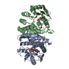
| |||||||||||||||||||||||||
| 2 | 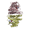
| |||||||||||||||||||||||||
| 3 | 
| |||||||||||||||||||||||||
| Unit cell |
| |||||||||||||||||||||||||
| Noncrystallographic symmetry (NCS) | NCS domain:
| |||||||||||||||||||||||||
| Details | The biological assembly is a dimer formed between chanis A and B and by chains C and D |
-Components
| #1: Protein | Mass: 29047.225 Da / Num. of mol.: 5 Source method: isolated from a genetically manipulated source Source: (gene. exp.) References: UniProt: P39610, phosphooxymethylpyrimidine kinase #2: Chemical | ChemComp-ADP /   Mass: 427.201 Da / Num. of mol.: 5 / Source method: obtained synthetically / Formula: C10H15N5O10P2 / Comment: ADP, energy-carrying molecule*YM Mass: 427.201 Da / Num. of mol.: 5 / Source method: obtained synthetically / Formula: C10H15N5O10P2 / Comment: ADP, energy-carrying molecule*YMHas protein modification | Y | |
|---|
-Experimental details
-Experiment
| Experiment | Method: X-RAY DIFFRACTION / Number of used crystals: 1 |
|---|
- Sample preparation
| Crystal | Density Matthews: 2.27 Å3/Da / Density % sol: 45.86 % |
|---|---|
| Crystal grow | Temperature: 290 K / Method: vapor diffusion, hanging drop / pH: 8.5 Details: 28% peg 4000, 0.17M sodium acetate trihydrate, 0.1M Tris HCL (pH 8.5), 10mM MgCl, 10mM ADP, VAPOR DIFFUSION, HANGING DROP, temperature 290K |
-Data collection
| Diffraction | Mean temperature: 100 K |
|---|---|
| Diffraction source | Source: ROTATING ANODE / Type: RIGAKU / Wavelength: 1.54 Å |
| Detector | Type: MARRESEARCH / Detector: IMAGE PLATE / Date: May 9, 2005 |
| Radiation | Monochromator: Osmic Varimax mirrors / Protocol: SINGLE WAVELENGTH / Monochromatic (M) / Laue (L): M / Scattering type: x-ray |
| Radiation wavelength | Wavelength: 1.54 Å / Relative weight: 1 |
| Reflection | Resolution: 2.8→94.916 Å / Num. all: 33604 / Num. obs: 33604 / % possible obs: 99.4 % / Observed criterion σ(F): 0 / Observed criterion σ(I): 0 / Redundancy: 3.7 % / Biso Wilson estimate: 50.3 Å2 / Rmerge(I) obs: 0.131 / Rsym value: 0.131 / Net I/σ(I): 5.7 |
| Reflection shell | Resolution: 2.8→2.95 Å / Redundancy: 3.8 % / Rmerge(I) obs: 0.475 / Mean I/σ(I) obs: 1.7 / Num. measured all: 18268 / Num. unique all: 4844 / Rsym value: 0.475 / % possible all: 99.9 |
- Processing
| Software |
| ||||||||||||||||||||||||||||||||||||||||||||||||||||||||||||||||||||||||||||||||||||||||||
|---|---|---|---|---|---|---|---|---|---|---|---|---|---|---|---|---|---|---|---|---|---|---|---|---|---|---|---|---|---|---|---|---|---|---|---|---|---|---|---|---|---|---|---|---|---|---|---|---|---|---|---|---|---|---|---|---|---|---|---|---|---|---|---|---|---|---|---|---|---|---|---|---|---|---|---|---|---|---|---|---|---|---|---|---|---|---|---|---|---|---|---|
| Refinement | Method to determine structure: MOLECULAR REPLACEMENT Starting model: 1UB0 Resolution: 2.8→12.87 Å / Cor.coef. Fo:Fc: 0.906 / Cor.coef. Fo:Fc free: 0.865 / SU B: 15.994 / SU ML: 0.317 / Isotropic thermal model: isotropic / Cross valid method: THROUGHOUT / σ(F): 0 / σ(I): 0 / ESU R Free: 0.441 / Stereochemistry target values: MAXIMUM LIKELIHOOD
| ||||||||||||||||||||||||||||||||||||||||||||||||||||||||||||||||||||||||||||||||||||||||||
| Solvent computation | Ion probe radii: 0.8 Å / Shrinkage radii: 0.8 Å / VDW probe radii: 1.2 Å / Solvent model: MASK | ||||||||||||||||||||||||||||||||||||||||||||||||||||||||||||||||||||||||||||||||||||||||||
| Displacement parameters | Biso mean: 31.436 Å2
| ||||||||||||||||||||||||||||||||||||||||||||||||||||||||||||||||||||||||||||||||||||||||||
| Refine analyze | Luzzati coordinate error obs: 0.39 Å | ||||||||||||||||||||||||||||||||||||||||||||||||||||||||||||||||||||||||||||||||||||||||||
| Refinement step | Cycle: LAST / Resolution: 2.8→12.87 Å
| ||||||||||||||||||||||||||||||||||||||||||||||||||||||||||||||||||||||||||||||||||||||||||
| Refine LS restraints |
| ||||||||||||||||||||||||||||||||||||||||||||||||||||||||||||||||||||||||||||||||||||||||||
| Refine LS restraints NCS | Dom-ID: 1 / Refine-ID: X-RAY DIFFRACTION
| ||||||||||||||||||||||||||||||||||||||||||||||||||||||||||||||||||||||||||||||||||||||||||
| LS refinement shell | Resolution: 2.8→2.869 Å / Total num. of bins used: 20
|
