 Movie
Movie Controller

[English] 日本語
 Yorodumi
- PDB-1xu7: Crystal Structure of the Interface Open Conformation of Tetrameri... -
+ Open data

- Basic information
| Entry | Database: PDB / ID: 1xu7 | ||||||
|---|---|---|---|---|---|---|---|
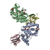
| Title | Crystal Structure of the Interface Open Conformation of Tetrameric 11b-HSD1 | ||||||
 Components Components | Corticosteroid 11-beta-dehydrogenase, isozyme 1 | ||||||
 Keywords Keywords | OXIDOREDUCTASE / 11b-HSD1 / SDR / dehydrogenase / hydroxysteroid | ||||||
| Function / homology |  Function and homology information Function and homology informationcortisol dehydrogenase (NADP+) activity / 11-beta-hydroxysteroid dehydrogenase (NADP+) activity / 11beta-hydroxysteroid dehydrogenase / 7beta-hydroxysteroid dehydrogenase (NADP+) / 7-beta-hydroxysteroid dehydrogenase (NADP+) activity / Glucocorticoid biosynthesis / steroid catabolic process / Prednisone ADME / steroid binding / lung development ...cortisol dehydrogenase (NADP+) activity / 11-beta-hydroxysteroid dehydrogenase (NADP+) activity / 11beta-hydroxysteroid dehydrogenase / 7beta-hydroxysteroid dehydrogenase (NADP+) / 7-beta-hydroxysteroid dehydrogenase (NADP+) activity / Glucocorticoid biosynthesis / steroid catabolic process / Prednisone ADME / steroid binding / lung development / NADP binding / endoplasmic reticulum membrane / protein homodimerization activity / membrane Similarity search - Function | ||||||
| Biological species |  Homo sapiens (human) Homo sapiens (human) | ||||||
| Method |  X-RAY DIFFRACTION / SYNCHROTRON / MAD / Resolution: 1.8 Å X-RAY DIFFRACTION / SYNCHROTRON / MAD / Resolution: 1.8 Å | ||||||
 Authors Authors | Hosfield, D.J. / Wu, Y. / Skene, R.J. / Hilger, M. / Jennings, A. / Snell, G.P. / Aertgeerts, K. | ||||||
 Citation Citation | Journal: J.Biol.Chem. / Year: 2005 Title: Conformational Flexibility in Crystal Structures of Human 11beta-hydroxysteroid dehydrogenase type I provide insights into glucocorticoid interconversion and enzyme regulation. Authors: Hosfield, D.J. / Wu, Y. / Skene, R.J. / Hilger, M. / Jennings, A. / Snell, G.P. / Aertgeerts, K. | ||||||
| History |
|
- Structure visualization
| Structure viewer | Molecule: MolmilJmol/JSmol |
|---|
- Downloads & links
-Download
| PDBx/mmCIF format | 1xu7.cif.gz | 226.9 KB | Display | PDBx/mmCIF format |
|---|---|---|---|---|
| PDB format | pdb1xu7.ent.gz | 183 KB | Display | PDB format |
| PDBx/mmJSON format | 1xu7.json.gz | Tree view | PDBx/mmJSON format | |
| Others |  Other downloads Other downloads |
-Validation report
| Arichive directory | https://data.pdbj.org/pub/pdb/validation_reports/xu/1xu7ftp://data.pdbj.org/pub/pdb/validation_reports/xu/1xu7 | HTTPS FTP |
|---|
-Related structure data


-Links
 PDBj
PDBj


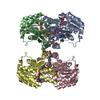
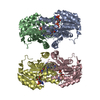
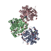
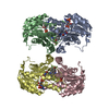
- Assembly
| Deposited unit | 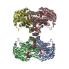
| ||||||||
|---|---|---|---|---|---|---|---|---|---|
| 1 |
| ||||||||
| 2 | 
| ||||||||
| 3 | 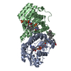
| ||||||||
| Unit cell |
| ||||||||
| Details | Biological tetramer |
-Components
| #1: Protein | Mass: 31836.875 Da / Num. of mol.: 4 / Mutation: C272S Source method: isolated from a genetically manipulated source Source: (gene. exp.) Homo sapiens (human) / Gene: HSD11B1, HSD11, HSD11L / Plasmid: pBAD-ThioE / Cell (production host): DH10b-Tir / Production host:  References: UniProt: P28845, 11beta-hydroxysteroid dehydrogenase #2: Chemical | ChemComp-NDP /   Mass: 745.421 Da / Num. of mol.: 4 / Source method: obtained synthetically / Formula: C21H30N7O17P3 Mass: 745.421 Da / Num. of mol.: 4 / Source method: obtained synthetically / Formula: C21H30N7O17P3#3: Chemical | ChemComp-CPS / 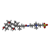  Mass: 614.877 Da / Num. of mol.: 6 / Source method: obtained synthetically / Formula: C32H58N2O7S / Comment: detergent*YM Mass: 614.877 Da / Num. of mol.: 6 / Source method: obtained synthetically / Formula: C32H58N2O7S / Comment: detergent*YM#4: Water | ChemComp-HOH / |  Mass: 18.015 Da / Num. of mol.: 486 / Source method: isolated from a natural source / Formula: H2O Mass: 18.015 Da / Num. of mol.: 486 / Source method: isolated from a natural source / Formula: H2O |
|---|
-Experimental details
-Experiment
| Experiment | Method: X-RAY DIFFRACTION / Number of used crystals: 1 |
|---|
- Sample preparation
| Crystal | Density Matthews: 2.6 Å3/Da / Density % sol: 52.64 % |
|---|---|
| Crystal grow | Temperature: 298 K / Method: vapor diffusion, hanging drop / pH: 7 Details: PEG , MES buffer, pH 7.0, VAPOR DIFFUSION, HANGING DROP, temperature 298K |
-Data collection
| Diffraction source | Source: SYNCHROTRON / Site: ALS  / Beamline: 5.0.3 / Wavelength: 1.3404, 1.3408, 1.3412, 1.2782 / Beamline: 5.0.3 / Wavelength: 1.3404, 1.3408, 1.3412, 1.2782 | |||||||||||||||
|---|---|---|---|---|---|---|---|---|---|---|---|---|---|---|---|---|
| Detector | Type: ADSC QUANTUM 4 / Detector: CCD / Date: Jun 1, 2003 | |||||||||||||||
| Radiation | Protocol: MAD / Monochromatic (M) / Laue (L): M / Scattering type: x-ray | |||||||||||||||
| Radiation wavelength |
| |||||||||||||||
| Reflection | Resolution: 1.8→20 Å / Num. all: 114558 / Num. obs: 114558 / % possible obs: 95.3 % / Observed criterion σ(F): 1 / Observed criterion σ(I): 1 / Rsym value: 0.046 / Net I/σ(I): 26.2 | |||||||||||||||
| Reflection shell | Resolution: 1.8→1.86 Å / Redundancy: 1.5 % / Mean I/σ(I) obs: 3 / Num. unique all: 9105 / Rsym value: 0.3 / % possible all: 76.2 |
- Processing
| Software |
| ||||||||||||||||||||||||||||||||||||||||||||||||||||||||||||||||||||||
|---|---|---|---|---|---|---|---|---|---|---|---|---|---|---|---|---|---|---|---|---|---|---|---|---|---|---|---|---|---|---|---|---|---|---|---|---|---|---|---|---|---|---|---|---|---|---|---|---|---|---|---|---|---|---|---|---|---|---|---|---|---|---|---|---|---|---|---|---|---|---|---|
| Refinement | Method to determine structure: MAD / Resolution: 1.8→20 Å / Cor.coef. Fo:Fc: 0.945 / Cor.coef. Fo:Fc free: 0.933 / SU B: 2.488 / SU ML: 0.078 / Cross valid method: THROUGHOUT / σ(F): 0 / σ(I): 0 / ESU R: 0.128 / ESU R Free: 0.116 / Stereochemistry target values: MAXIMUM LIKELIHOOD
| ||||||||||||||||||||||||||||||||||||||||||||||||||||||||||||||||||||||
| Solvent computation | Ion probe radii: 0.8 Å / Shrinkage radii: 0.8 Å / VDW probe radii: 1.4 Å / Solvent model: BABINET MODEL WITH MASK | ||||||||||||||||||||||||||||||||||||||||||||||||||||||||||||||||||||||
| Displacement parameters | Biso mean: 17.86 Å2
| ||||||||||||||||||||||||||||||||||||||||||||||||||||||||||||||||||||||
| Refinement step | Cycle: LAST / Resolution: 1.8→20 Å
| ||||||||||||||||||||||||||||||||||||||||||||||||||||||||||||||||||||||
| Refine LS restraints |
| ||||||||||||||||||||||||||||||||||||||||||||||||||||||||||||||||||||||
| LS refinement shell | Resolution: 1.8→1.843 Å / Total num. of bins used: 20 /
|
